At the Lotus Gemology laboratory in Bangkok, we often get parcels of relatively uniform stones. But sometimes it is in this routine testing that we uncover surprises.
Introduction
At Lotus Gemology, our Bangkok-based gem testing laboratory, we only test for two types of stones: corundum (ruby & sapphire) and spinel. As a result we often get parcels of stones with similar attributes. This means that sometimes we can test twenty or thirty untreated Mozambique rubies in a row, for example. Often it is in the routine testing of these seemingly uniform parcels that we uncover surprises.
We recently got one such lot of 11 light yellow sapphires, ranging from approximately 2–5 carats. This is a standard, unremarkable submission for our lab.
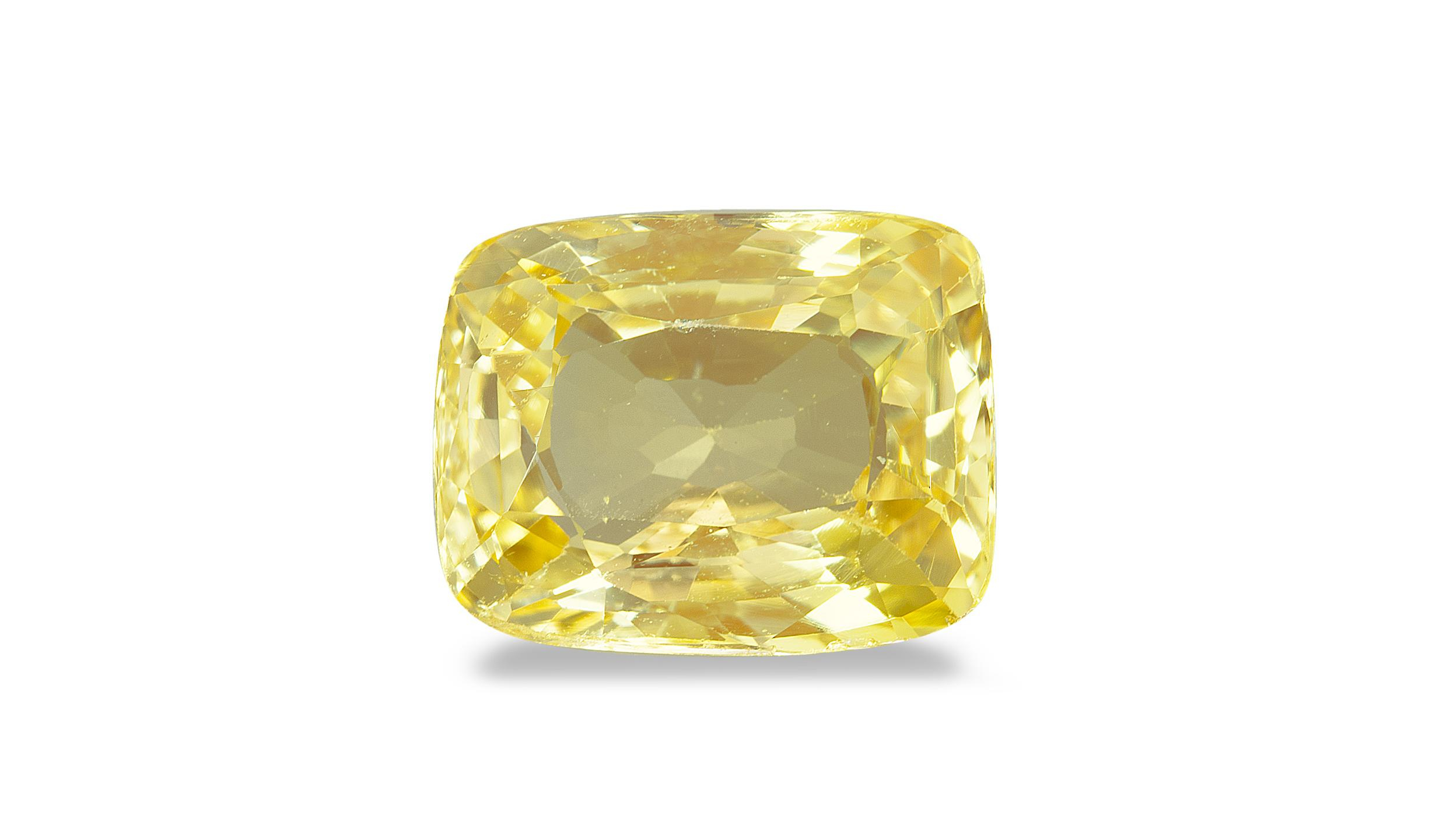 Figure 1. The 4+-carat yellow stone that is the subject of this report. Photo: Maitree Petchloun
Testing
Appearance
The appearance of the 11 stones in the lot was relatively homogenous. All displayed a pleasant yellow hue, with rich to pastel saturation, and all had a medium-light to light tone. Even the cutting was similar, with most stones in the parcel cut with a modified brilliant crown and either fancy or step cut pavilion. At Lotus, lots of yellow sapphires with this appearance are common.
UV Fluorescence
After noting the usual identifying features of the stones, including weight, measurements, and appearance, we checked the stones’ reactions in fluorescent lighting.
10 of the yellow stones in the lot displayed similar fluorescence patterns with medium yellow to orange fluorescence across the stone in the longwave light, and weak to medium orange fluorescence in the shortwave light.
In contrast, one stone had a medium red-orange fluorescence in the long wave and weak reddish-orange fluorescence in the shortwave. Under both wavelengths, the fluorescence of our subject was markedly redder than that of the other yellow sapphires in the lot. This was the first sign of something unusual.
Visible Spectrum
Usually in natural yellow sapphire, as was the case with most of the stones submitted in this parcel, a weak Fe (iron) spectrum can be seen. However, the stone with the reddish fluorescence did not display a diagnostic spectrum.
This could be cause for concern, yet detecting a weak iron spectrum using the direct-vision spectroscope can be tricky for many gemologists. (To learn more, see our article “Gem Testing with the Spectroscope.”) In fact, a weak line at about 455 nm has even been reported in synthetic flame fusion corundum (Koivula, 2005). Thus, more testing had to be done.
Infrared Spectrum
When tested with the FTIR (Fourier transform infrared spectroscopy), nine of the 11 stones submitted displayed a weak to strong peak at 3160. Two spectra did not display diagnostic features, including the one associated with our subject.
It is a little suspicious that these two spectra stood out from the rest, but that is not diagnostic evidence when viewed alone. Because of factors such as differences in cutting, some stones may not display a strong signal that provides a clear, diagnostic spectrum.
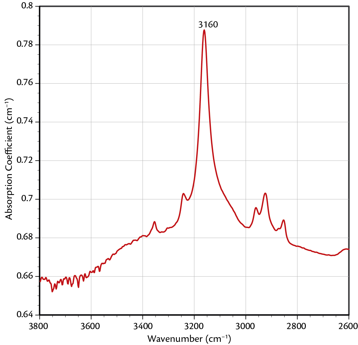 Figure 2. The above spectrum was shows a strong 3160 peak typical of the natural sapphires in the parcel.
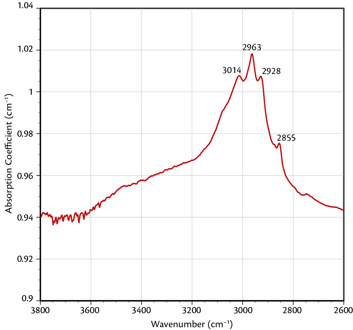 Figure 3. This is the spectrum of the subject of this report. It did not show any diagnostic features at 3160 nm.
Inclusions
Most of the stones in the lot displayed inclusions typical of untreated Sri Lankan sapphire. These include angular zoned clouds of exsolved particles, angular growth zoning, partially healed fissures (fingerprints), euhedral negative crystals, and small uraninite crystals.
The stone in question was almost entirely free of inclusions. The only inclusions we could find using dark field lighting in the microscope were a few shallow fissures and a series of small pinpoint dots near the culet. Because of their small size, it was hard to tell whether these dots were exsolved particles, tiny crystals, or gas bubbles.
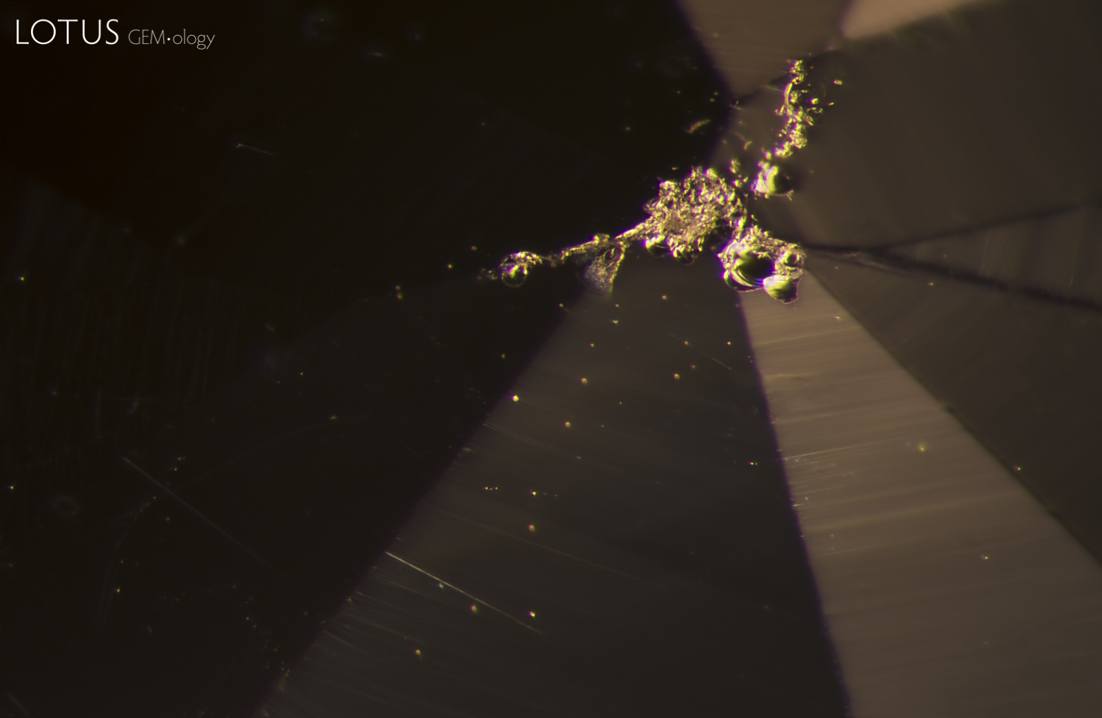 Figure 4. Small, bright scattered dots are visible under the culet of this stone. Upon further testing, we determined that they are gas bubbles, a common feature of Verneuil synthetic corundum. (The abrasions at the intersection of the facet edges are small chips at the culet.) Photo: E. Billie Hughes.
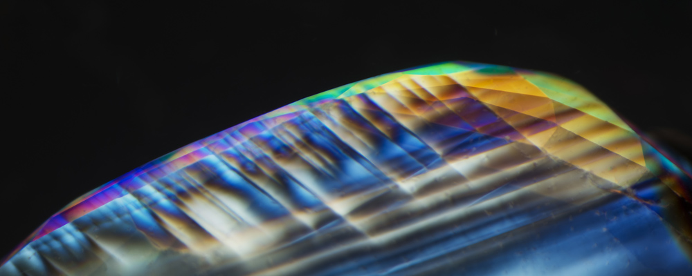
To do further testing, we immersed the stone in iodomethane (methylene iodide) under crossed polars. Then we saw iridescent rainbow lines dancing across the stone. With a cursory glance this could be mistaken for angular growth zoning. However, after rotating the stone in different directions, we could see that these features actually followed parallel twinning that intersected at 60° or 120° angles, termed Sandmeier-Plato twinning. These twin planes form parallel to the hexagonal prism and are a diagnostic feature of flame fusion synthetic corundum (Eppler, 1964; Gübelin, 1986, 2005; Hughes, 2017).
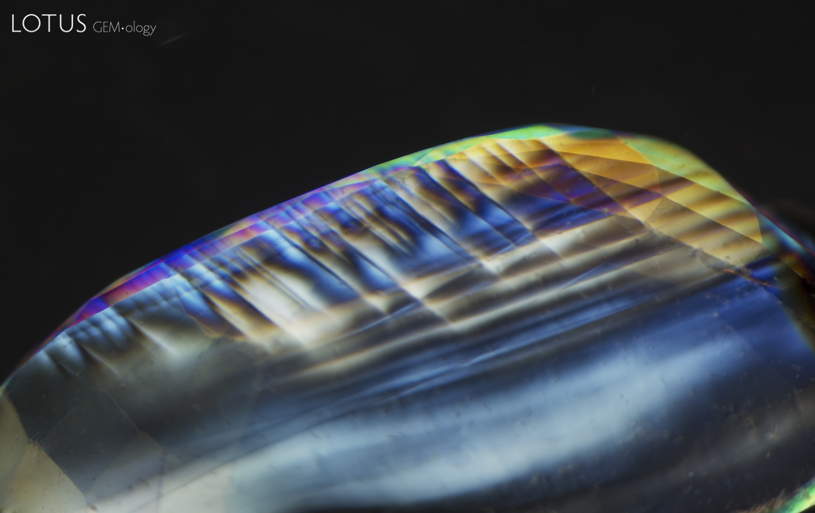 Figure 5. Sandmeier-Plato twinning is a type of polysynthetic twinning parallel to the hexagonal prism. These lines are proof of Verneuil synthetic origin. Photo taken under crossed polars while the stone was immersed in iodomethane (methylene iodide). Photo: E. Billie Hughes.
Chemistry
We also analyzed the chemical composition of the stone in question using EDXRF (energy dispersive x-ray fluorescence) (Table 1).
Two features stood out that confirmed our suspicions. One was the complete lack of Ga (gallium) at 0.000%. The other was the extremely low level of Fe (iron) at 0.0010%. In natural sapphire we would expect to see higher levels of both of these elements (Muhlmeister, 1998; Hughes, 2016). This suggests that the specimen is synthetic.
| Element | Weight % |
|---|---|
| Al2O3 | 99.9956 |
| Cr2O3 | 0.0033 |
| Fe2O3 | 0.0010 |
| Ga2O3 | 0.0000 |
Conclusion
It is not any single gemological test that tells us the true nature of a stone. Rather, it is the combination of evidence from a series of tests, which, when seen as a whole, helps us form a conclusion.
In this instance, the unusual red fluorescence, coupled with a lack of diagnostic visual and infrared spectra, raised our suspicions about the nature of the stone. These suspicions were then confirmed when we found gas bubbles and Sandmeier-Plato twinning with microscopic observation. Chemical analysis further solidified the evidence that this seemingly typical yellow sapphire was actually a flame fusion synthetic, hidden among natural stones.
![]()
About the Author
E. Billie Hughes is Co-Founder and Managing Director of Lotus Gemology. She oversees the company's day-to-day operations while continuing gemological research and laboratory work. After graduating from UCLA in 2011, Billie became a Fellow of the Gemmological Association of Great Britain (FGA) in 2013. Her research focuses on ruby and sapphire, including low-temperature heat treatment, and she has authored and co-authored articles in leading gemological journals. An accomplished field gemologist, she has traveled to gem deposits around the world, including nearly every major ruby and sapphire locality.
Billie is an internationally recognized educator who has lectured for trade organizations, museums, and luxury jewelry houses. She has collaborated extensively with Van Cleef & Arpels on educational programs and lectures. An award-winning photographer and photomicrographer, her images have received honors in the Nikon Small World and Gem-A competitions and have appeared in publications including National Geographic and Forbes. She is also the creator of Hyperion, Lotus Gemology's online inclusion database, reflecting her commitment to making gemological knowledge more accessible.
Billie developed an interest in gemstones from an early age, accompanying her parents on expeditions to mines and gem-producing regions around the world. That lifelong passion for fieldwork, laboratory research, education, and photography continues to shape her work at Lotus Gemology today.
Notes
This article first appeared in Gem Market News, companion to the GemGuide, July/August 2017, Vol. 36, Issue 4, pp. 18–20, published by Gemworld International, Inc., Glenview, Illinois. Reprinted with permission.
This article also appeared in The Journal of the Gemmological Assoication of Hong Kong, 2017, Volume XXXVIII, pp. 47–49.
![]()
Acknowledgements
The author would like to thank Richard W. Hughes, Elise Skalwold, and Patharaphum Sudprasert for their help.
References & further reading
- Eppler, W.F. (1964) Polysynthetic twinning in synthetic corundum. Gems & Gemology, Vol. 11, No. 6, Summer, pp. 169–175.
- Gübelin, E.J. and Koivula, J.I. (1986) Photoatlas of Inclusions in Gemstones. Zürich, Switzerland, ABC Edition, revised Jan., 1992
- Gübelin, E.J. and Koivula, J.I. (2005) Photoatlas of Inclusions in Gemstones, Volume 3. Basel, Switzerland, Opinio Publishers, 672 pp.
- Hughes, E. B., Chankhantha, C. et al. (2016) Padparadscha or Pretender: An Unusual Pink-Orange Sapphire. The Australian Gemmologist, Vol. 25, Nos.11–12, pp. 389–392.
- Hughes, R.W., Manorotkul, W. & Hughes, E. B. (2017) Ruby & Sapphire: A Gemologist’s Guide. Lotus Publishing, Bangkok, 816 pp.
- Koivula, J.I. & Hughes, R.W. (2015) Gem testing with the spectroscope. Lab World Magazine, Vol. 04, No. 04, May–July, pp. 05–08
- Muhlmeister, S., Fritsch, E. et al. (1998) Separating natural and synthetic rubies on the basis of trace-element chemistry. Gems & Gemology, Vol. 34, No. 2, Summer, pp. 80–101
![]()