Results 61 – 77 of 77 sets.


A. One of the oldest gem frauds is the doublet, where a thin slice of a natural gem is attached to a larger piece of stone (typically synthetic or glass, but could also be natural). In this case, a thin slice of natural green sapphire is fused to a piece of synthetic ruby. Due to the thinness of the top piece, the color difference cannot be seen from the top, but it is unmasked in the microscope in immersion, as shown here. In rare cases, where a gem broke during cutting, the two pieces may be glued back together. This is not a doublet, but a “repaired” stone.
B. With all assembled stones, the key to identification is to locate the separation plane between the two pieces. It is almost always completely flat (due to polishing) and must continue unbroken around the entire stone. The separation plane is usually at the girdle.
C. When viewed from the top, flat gas bubbles can be found trapped in the separation plane. This stone’s natural crown also shows clouds of partially dissolved silk with internal diffusion, suggesting the top piece was heat treated. In Thailand, borax is often used in place of cement for producing corundum doublets. The borax dissolves the surfaces of the two pieces, fusing them together after cooling. This is similar to garnet-and-glass doublets, where the garnet is fused to the glass with heat. Fusion produces better doublets than using a glue, as cements may deteriorate with time.
D. This triplet is made by sandwiching two pieces of glass around a colored cement layer, where the colored cement gives color to the entire stone.


A. A prominent, intricate fingerprint pattern consisting of interconnected communication tubes extending from a mineral inclusion nucleus serves to confirm that this Sri Lankan spinel is natural. The form taken by the tubes reveals the isometric symmetry of the underlying spinel. Note the subtle differences in the healing pattern of this spinel relative to the previous corundum images.
B. Web-like secondary (’healed’) fingerprint in a natural spinel. Note how the healing pattern differs from that in corundum, due to spinel’s isometric structure.


A. In rubies from the Thailand/Cambodia border deposits, thin “two-phase” liquid films are a common site, forming in the plane of the basal pinacoid. These are generally hexagonal or triangular in shape with a bubble-like appearance in the center. Some may have hexagonal fingerprints around them.
B. Thin film inclusions in a ruby from the Thai/Cambodian border deposits. Note how the appearance changes dramatically depending on whether the inclusions are seen in transmitted light (upper left) versus reflected light (lower right).
C. Thin film inclusions in a ruby from the Thai/Cambodian border deposits. These form in the basal plane, perpendicular to the c-axis.
D. Diagram showing the orientation of thin film inclusions in Thai/Cambodian rubies. They form in the basal plane, perpendicular to the c-axis. In transmitted light, these inclusions may sometimes be mistaken for gas bubbles in synthetic corundum or glass.


A. In order to make synthetic corundums look more natural, dealers sometimes heat the rough and then cool it rapidly. This creates a fine honeycomb network of fissures. After cracking, the gem is then heat-treated with a flux, causing the fissures to heal. Upon first glance, the healed fissures resemble a natural ruby that has been heated, but closer observation reveals curved striae, proving that the stone is a quench-crackled synthetic ruby.
B. This Verneuil ruby synthetic was quench crackled and then heated with a flux to heal the fissures. Note the “honeycomb” pattern of the fissures, which is a telltale sign of quench crackling.
A. The most diagnostic inclusions in emeralds from Colombia are the three-phase inclusions. These are jagged-edged negative crystals filled with a liquid, a gas and a cube-shaped crystal of halite (sodium chloride or rock salt). However, three-phase inclusions may also be found in emeralds from Afghanistan, China (Davdar) and elsewhere.
B. This fascinating multiphase inclusion shows an unusual sawtooth pattern on the long edges. Many of the cavities contain liquid, gas and as many as four different solid phases.
C. Within this Afghan emerald, numerous cavities contain liquid, gas and as many as four different solid phases.
D. In contrast, emeralds from Zambia’s important Kafubu district display negative crystals in a prismatic pattern with a two-phase filling.
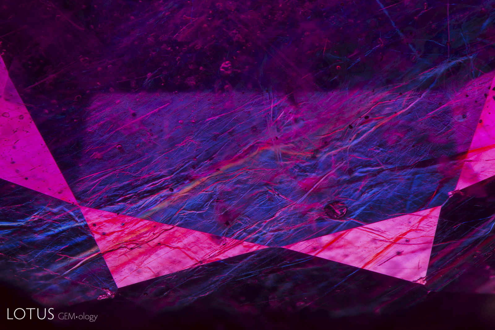
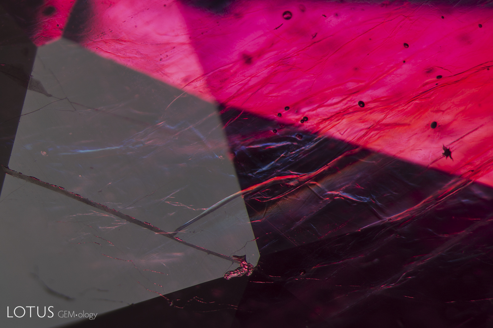
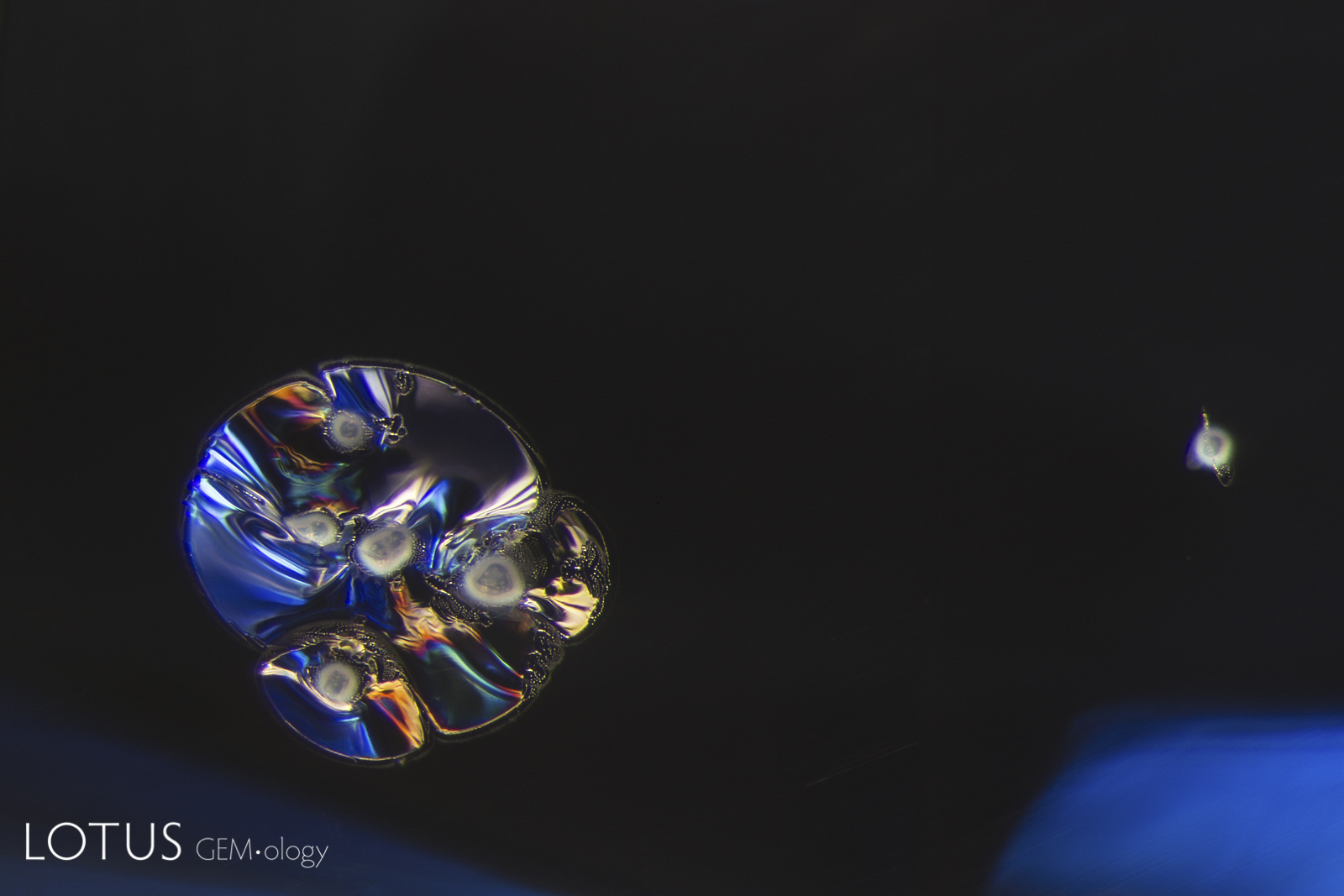
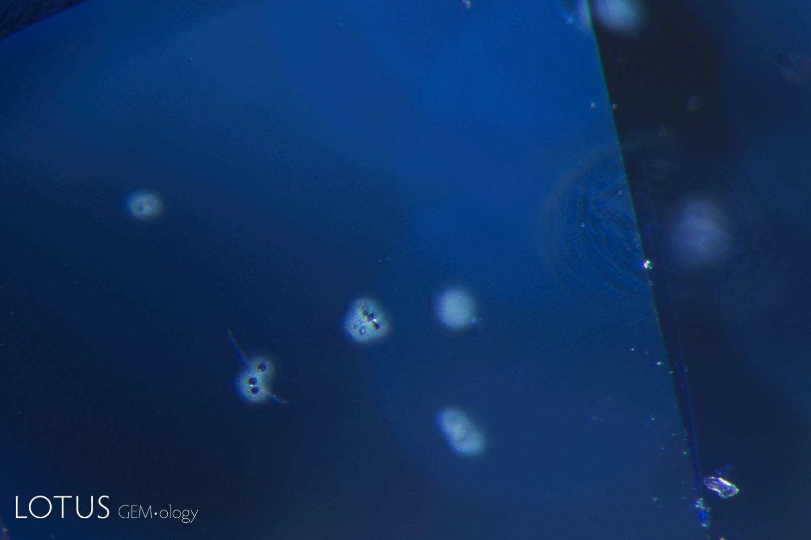
A. Complex fluid inclusions like these are occasionally found in spinel from Burma’s Mogok Stone Tract. The yellowish areas are a sulfur-rich liquid.
B. Another example of a sulfur-rich fluid inclusion in a Mogok spinel.

A. Exsolved rutile silk in a Sri Lanka sapphire. At high temperatures, crystals have more defects and a more-expanded lattice, and thus are better able to absorb impurities. As the crystal cools, defects are reduced. This may force impurities to crystallize out. But because of the constraints placed on their movement by the solid host, impurity atoms are unable to travel large distances. Therefore, rather than forming large crystals, they migrate short distances to form multitudes of tiny needles, plates and particles, along the directions in the host where space permits. This example not only shows rutile (TiO₂) needles, but the large example in the center features a daughter crystal at the broad end. The rutile actually forms small twinned crystals, as evidenced by the re-entrant angles at the broad end.
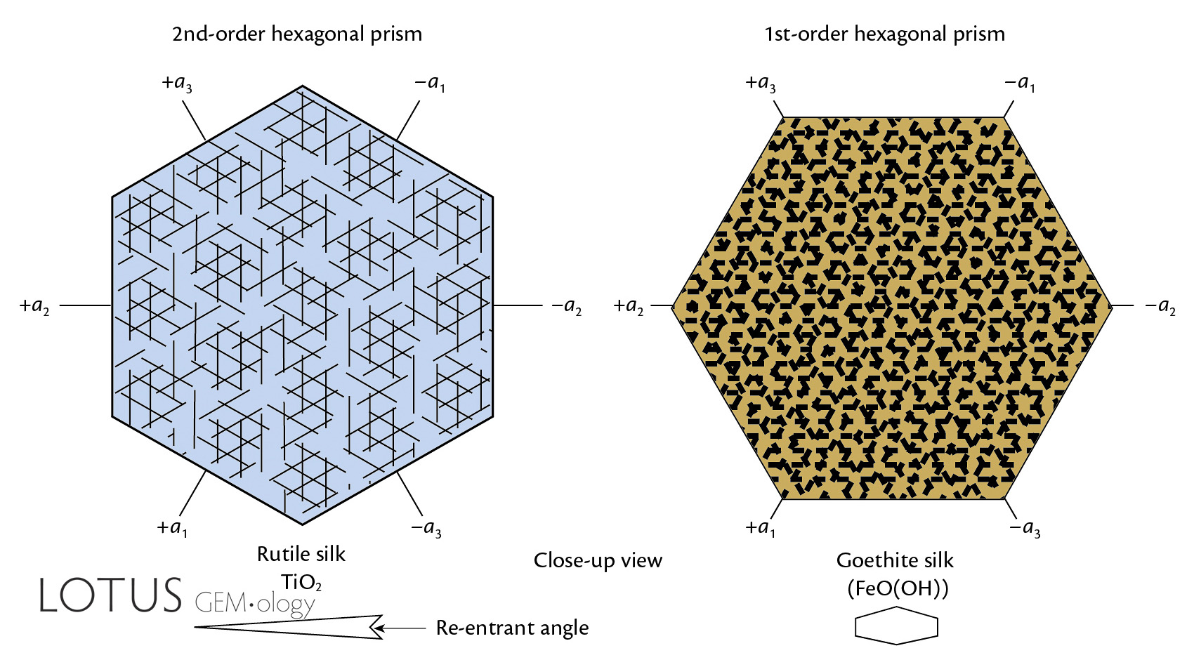
B. Silk in natural corundum is exsolved along three directions (parallel to the hexagonal prism) in the basal plane, meeting at 60/120°. Left: Rutile silk forms parallel to the faces of the second-order prism, and tends to occur as dart- or arrow-shaped twins with tiny reentrant angles at the broad end. Rutile silk is commonly found in corundum from a variety of sources, particularly Myanmar and Sri Lanka. At right, Fe-rich silk forms parallel to the faces of the first-order prism, and tends to be more platy (rather than slender needles). Fe-rich silk is common in Fe-rich sapphires, particularly those from Thailand, Australia and Kenya. Some Thai black-star sapphires (from Khao Ploi Waen and Bang Kha Cha) may contain both Fe-rich and rutile silk, and thus can be cut into 12-rayed star stones.
C. In sapphires that are rich in iron, the Fe and/or titanium may unmix in the form of “hairy” hematite/ilmenite/goethite silk, as shown in this “gold sheen” sapphire from Kenya.
D. When cut with the c-axis perpendicular to the silk layers, rubies and sapphires with silk inclusions can display a “star” effect, a phenomenon known as asterism. This example is rutile silk in a Myanmar ruby.


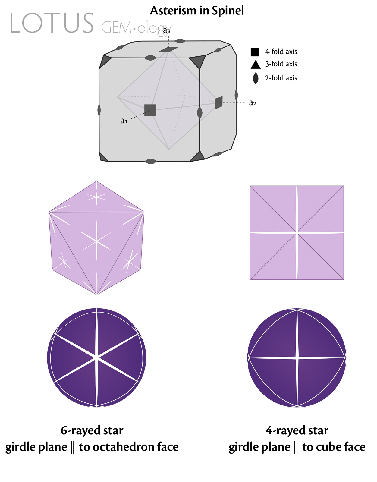
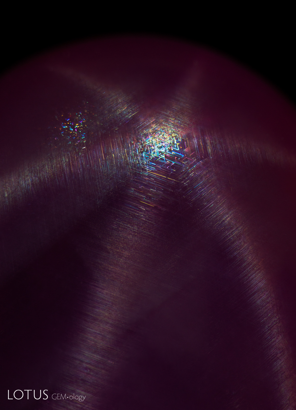
A. Fine silk creates a six-rayed star in this spinel. Spinel has the ability to display six- or four-rayed stars, depending on how the stone is oriented during cutting. Six-rayed stars are seen if the cabochon base is parallel to the octahedron face, while four-rayed stars occur when the base is parallel to a cube face. The silk inclusions in star spinel have been reported as being rutile, sphene (titanite), or sillimanite
B. In addition to having six-rayed stars, spinels like this one can also display four-rayed stars if the base is parallel to a cube face.
C. In star spinels, the exsolved silk unmixes parallel to the edges of the octahedron faces. Six-rayed stars are seen if the cabochon base is parallel to the octahedron face, while four-rayed stars occur when the base is parallel to a cube face.
A. The vivid blue and orange flashes seen in this stone, produced by glass-filled fissures, clearly identify it as a lead-glass hybrid ruby. This treatment involves cleaning low-grade ruby with acids to clean out secondary deposits in the fissures. After cleaning, the ruby’s fissures are then impregnated with a high refractive-index glass to mask the fissures.
B. Reflecting light from the surface of this stone reveals a difference in luster where the fissures have been filled with glass. Using dark field illumination we can also see bright flashes of color, confirming that this stone is a glass hybrid ruby. This example emphasizes the importance of looking at each stone in different lighting conditions.
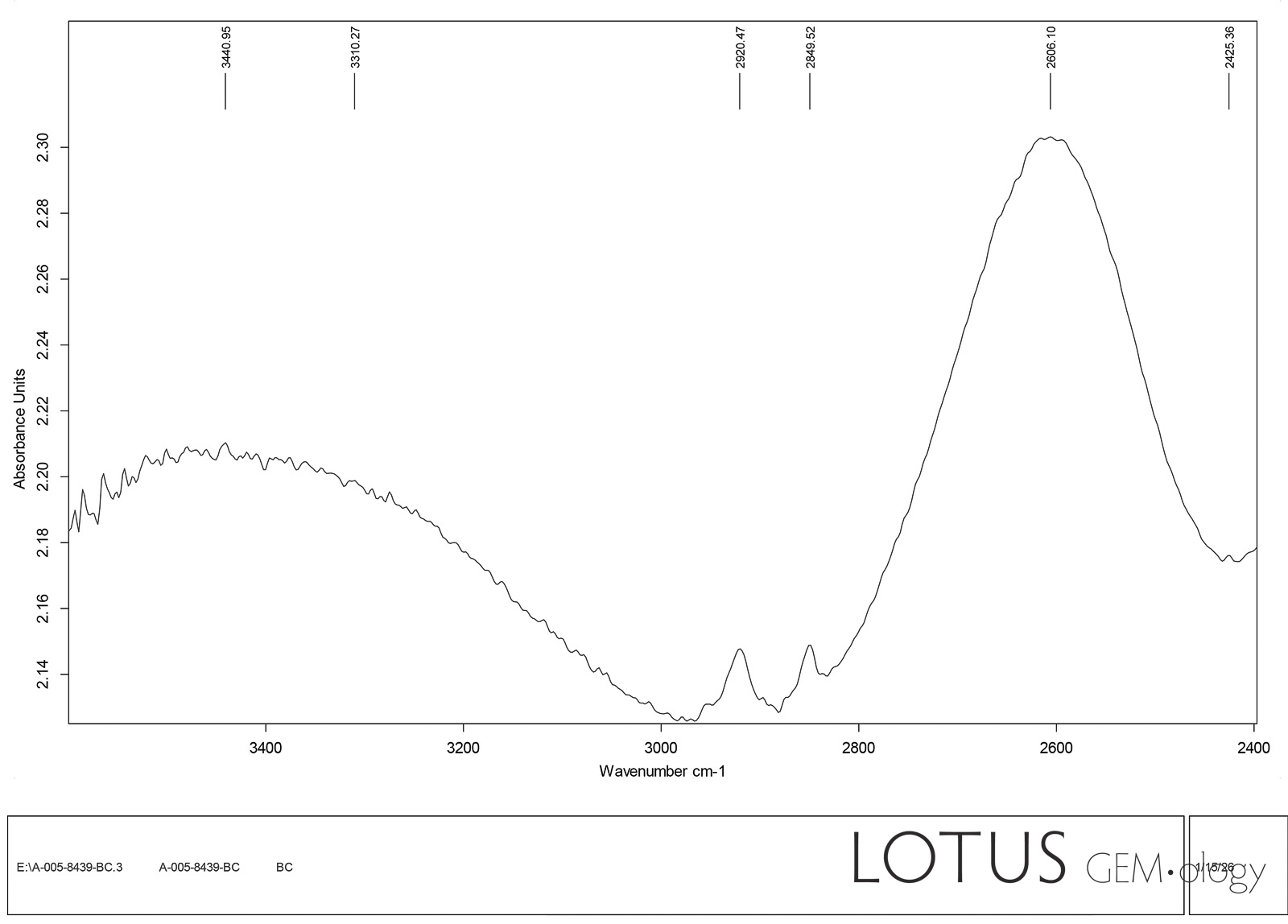
C. This glass-hybrid ruby displays a characteristic peak from 2500–2800 in the infrared, thus separating it from natural ruby, which will not display this peak.
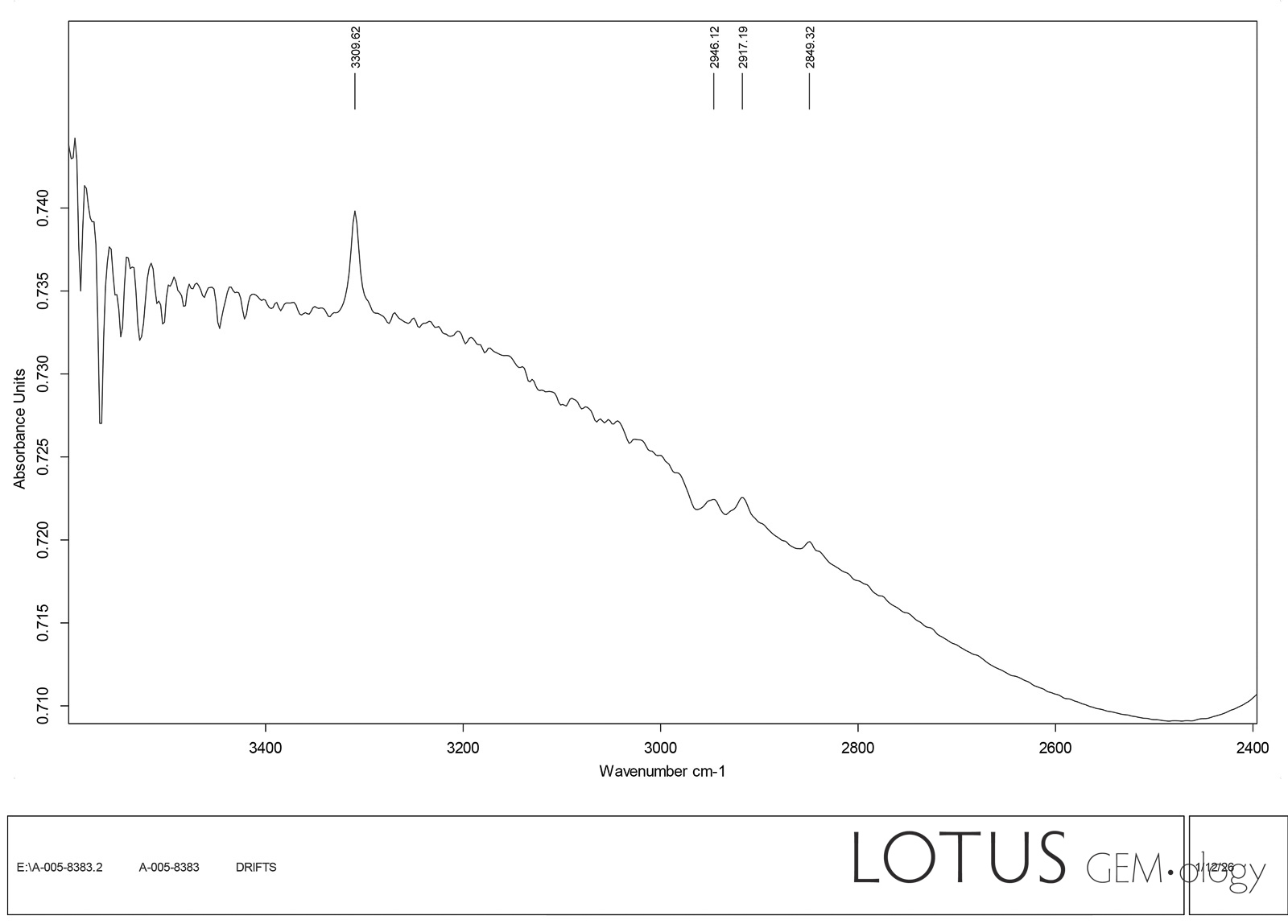
D. Contrast the infrared spectrum of the glass-filled ruby at left (C) with the natural ruby (D), showing that the spectra easily distinguish glass-filled from natural ruby.
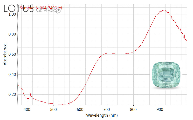
A. The UV-Vis-NIR spectrum is a fast and effective way of separating copper-bearing (’cuprian’) and non-cuprian tourmaline. In this blue-green tourmaline, the major chromophore is copper, as shown by the major peak at about 910 nm.
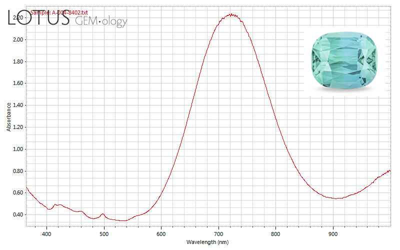
B. This pattern, with a large peak at about 710 nm, shows that this stone is not colored by copper. But if you compare the colors of the two gems (Gem A vs. Gem B), you can see that a virtually identical color is possible without copper. The marketplace pays a far higher price for the cuprian vs. the non-cuprian tourmaline. Why? To paraphrase the Irish rock band U2, the gem market “moves in mysterious ways.”
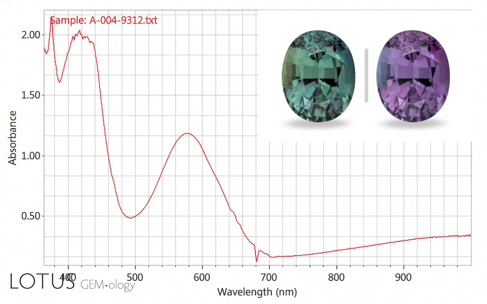
A. Alexandrite chrysoberyl is normally colored by the Cr3+ ion. This creates transmission in the blue-green and deep red ends of the spectrum, separated by a large absorption band in the yellow at about 580 nm. Because our eyes are more sensitive to green light, balanced daylight causes the color to appear greenish. In contrast, incandescent lights are extremely poor in the short wavelengths and strong in the yellow, orange and red wavelengths. This tips the balance, making them appear purple to red. The height of the absorption band in the yellow determines the strength of the color change.
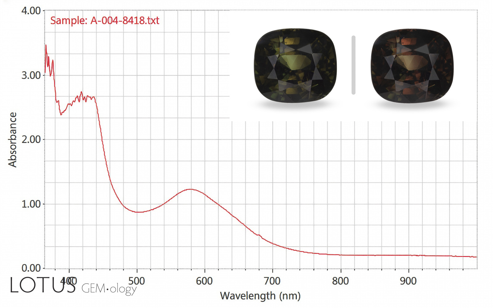
B. In this example, the alexandrite contains far more Fe than Cr. This produces a flatter absorption curve and thus a much weaker change-of-color.
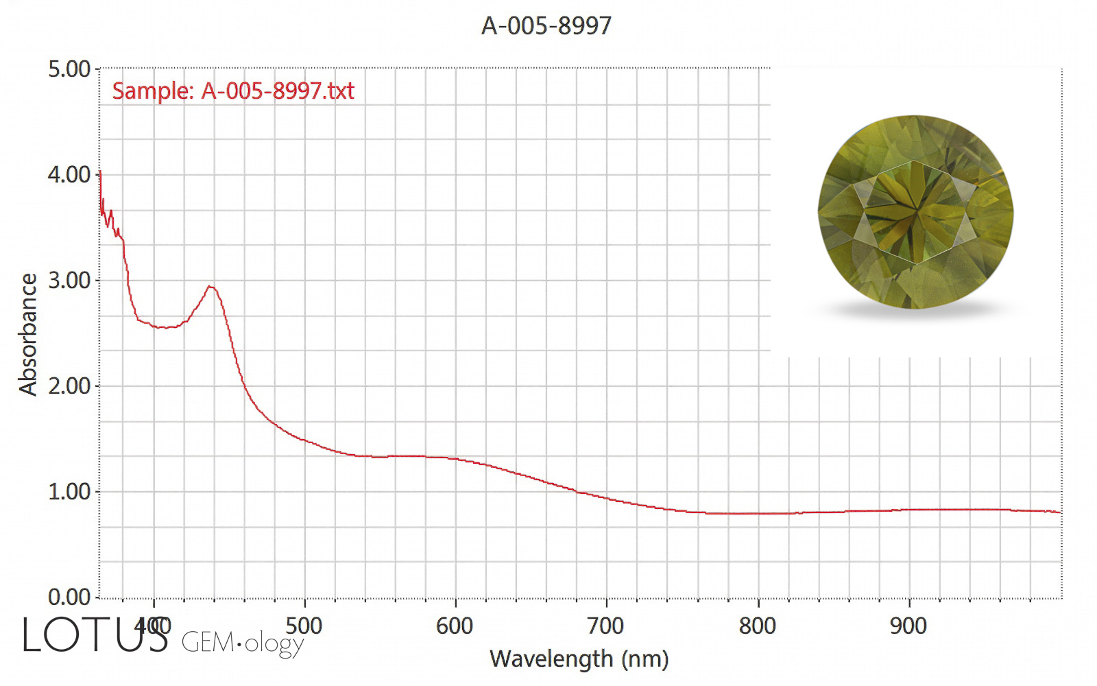
C. In gems with just a trace of Cr, the 580 band almost entirely disappears. The result is no color change.
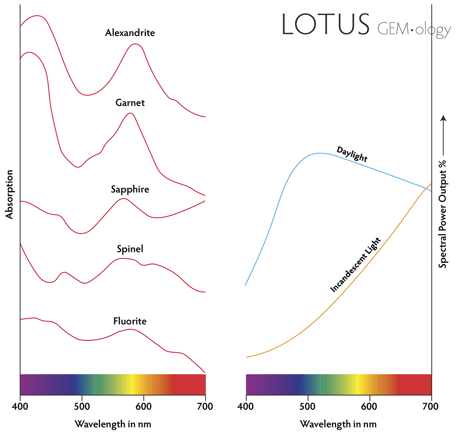
D. The absorption spectra of five different gems that display a color-change effect. Those that show the greatest difference between transmission valleys in the blue-green and red and the absorption peak in the yellow will show the strongest change of color (alexandrite and garnet). At right are the spectral power outputs of daylight vs. incandescent light. Daylight is relatively balanced, but incandescent light is much richer in long wavelengths. Illustration: Richard Hughes/Lotus Gemology (after Gübelin & Schmetzer, 1982).
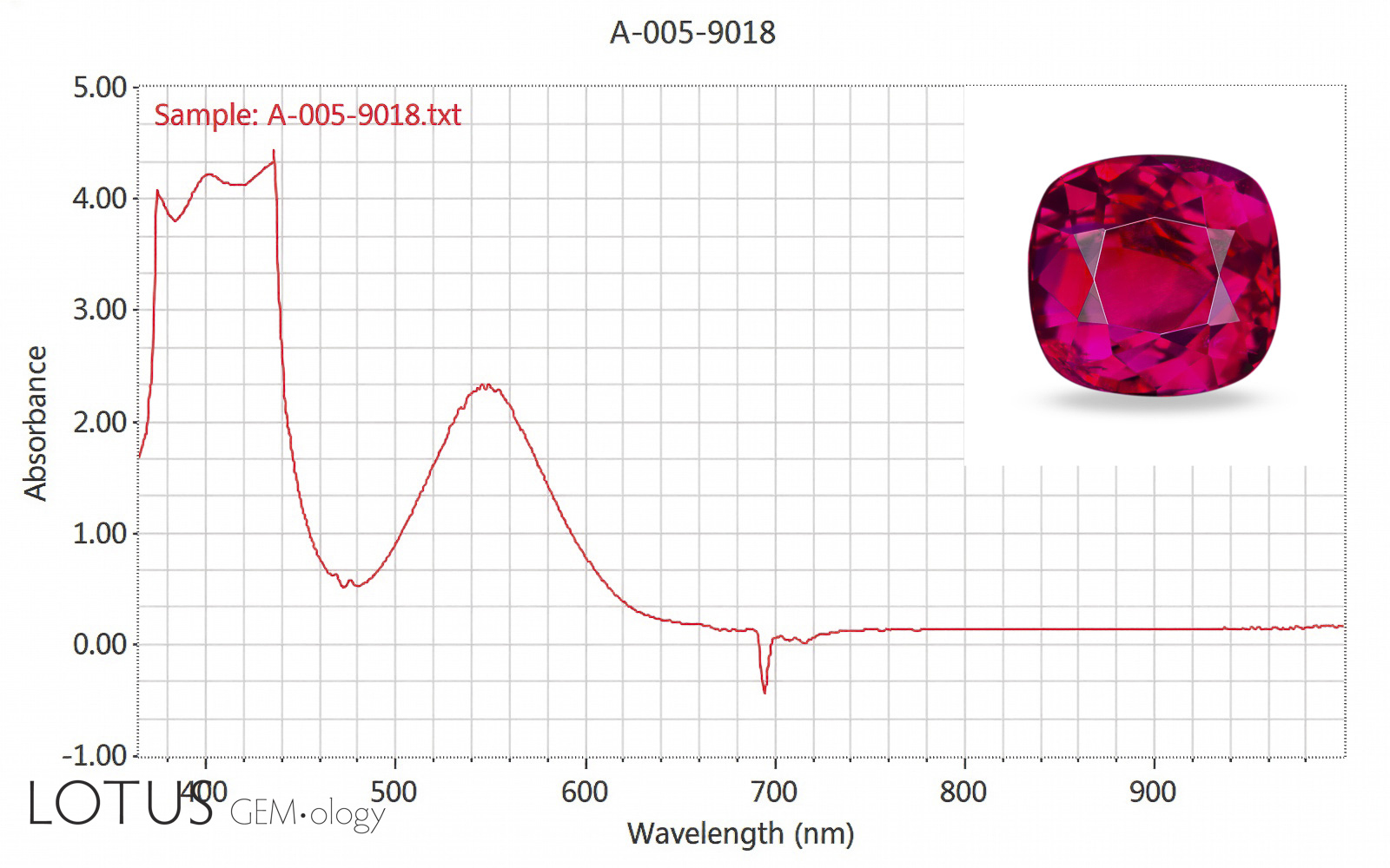
A. Note the three spectra in this set. All display the same basic absorption pattern of transmission in the blue-green and red and a large absorption band between the two. In ruby, that band is centered at about 550 nm. Ruby transmits a narrow area in the blue and lots of the red, giving ruby a slightly purplish red color. Note that corundum is Al₂O₃ and crystallizes in the trigonal system.
B. Chrysoberyl is BeAl₂O₄ and it crystallizes in the orthorhombic system. When Cr3+ substitutes for Al3+ in alexandrite, the slightly different composition and structure shifts the spectrum slightly to the longer wavelengths, producing a gem with a color that sits on the fence between the red of ruby and green of emerald. This change-of-color (color inconstancy) effect is highly prized in the gem market.
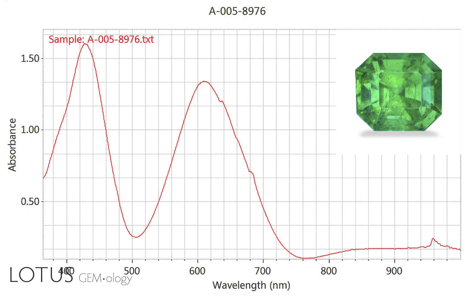
C. Beryl is Be₃Al₂(SiO₃)₆ and it crystallizes in the hexagonal system. When Cr3+ (or V3+) substitutes for Al3+ in emerald, it moves the entire absorption pattern further to the longer wavelengths, producing the classic green color of emerald. The reason for the differences in the three spectra has to do with the electron clouds. Due to the differences in composition and structure of each mineral, light is affected slightly differently. Note also that there may be absorption differences with direction (termed ‘pleochroism’) in non-isometric (doubly refractive) minerals.
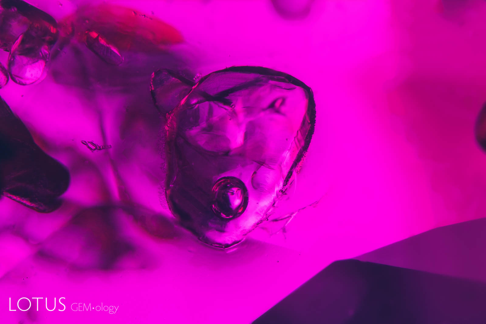
A. This cluster of melted crystals with a frosty “snowball” appearance and glassy discoid around them is a clear sign that their sapphire host has been heat treated.
B. Heat-altered “snowball” like crystals in a heat-treated sapphire from Sri Lanka. These inclusions are probably zircon.
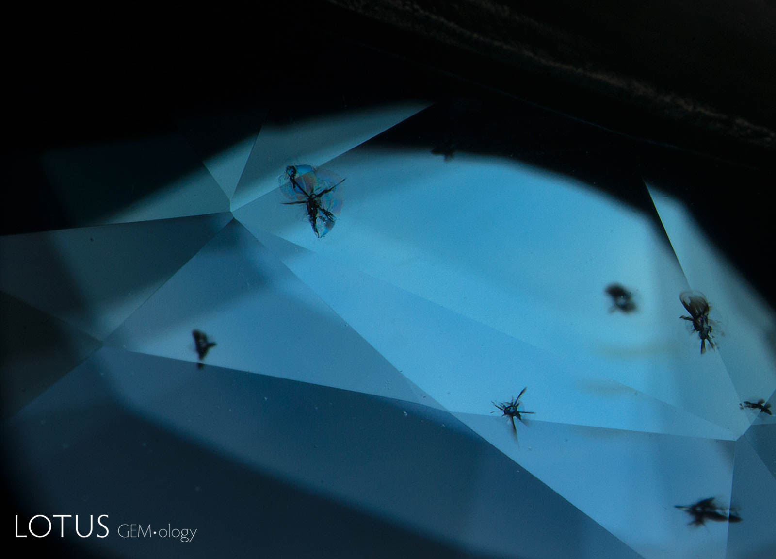
C. Resembling small flying insects, zircon crystals with radiation-induced stress halos are a common feature in Ceylon sapphires. This is what they look like before heat treatment.

A. This negative crystal in a Madagascar ruby displays a mobile bubble. Since the negative crystal had no opening to the surface, an oil-filled cavity could be safely ruled out. Such inclusions are usually liquid and gaseous carbon dioxide. Since they do not survive temperatures beyond a few hundred degrees centigrade, they generally constitute proof of natural, unheated origin.
B. Changing the position of the gem causes the bubble to move. Such fluid-filled cavities (generally filled with liquid and gaseous CO2) cannot withstand heat treatment and thus are proof of natural origin.
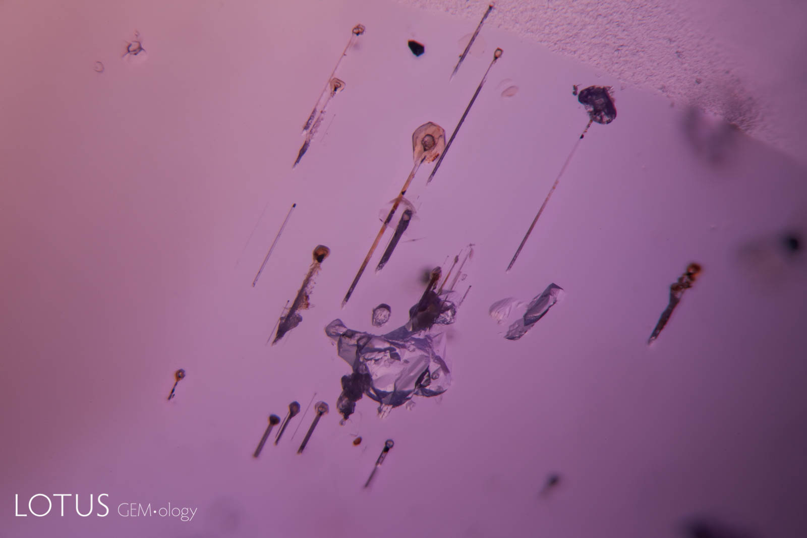
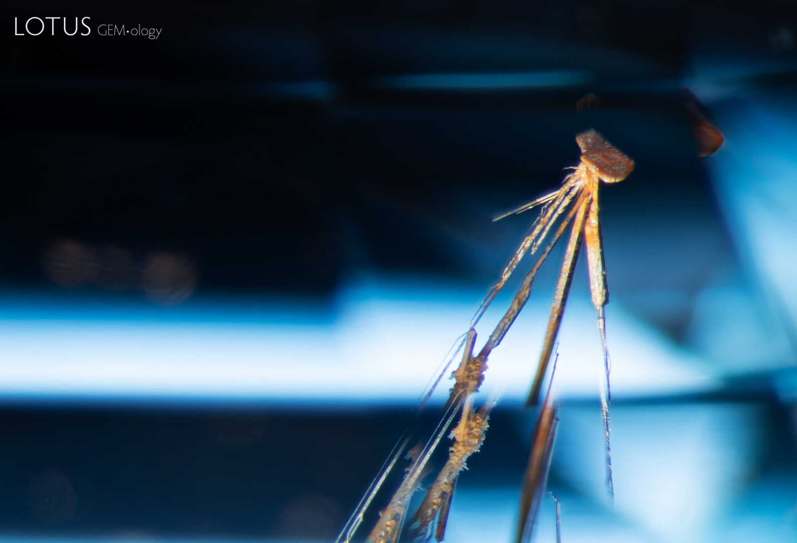
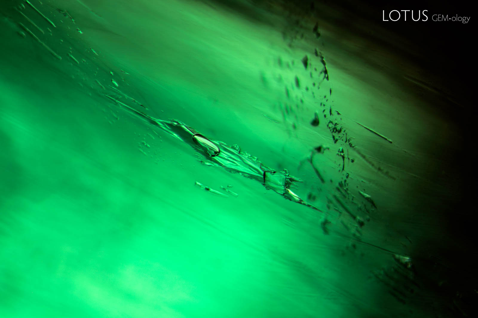
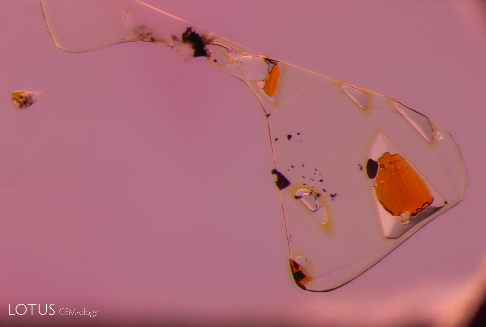
A. Like balloons on a string, these unusual crystals of what may be apatite with attached growth tubes, along with a negative crystal, create a beguiling scene in this purple star sapphire from Sri Lanka.
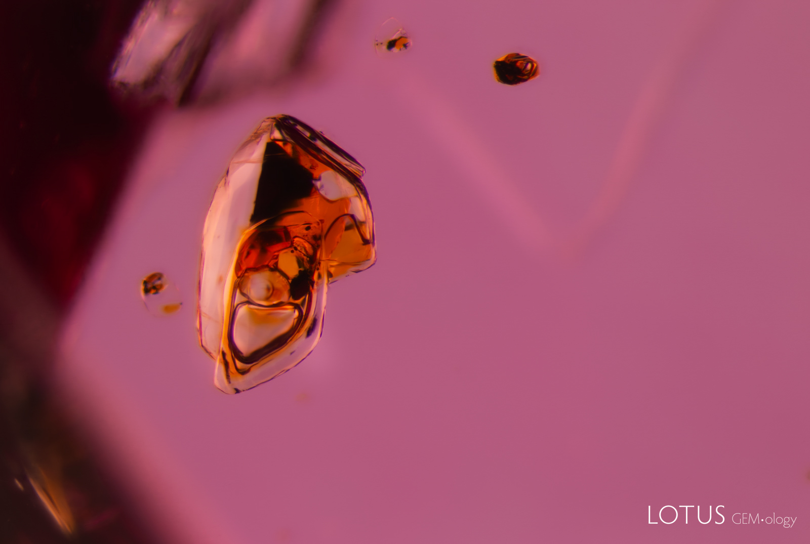
B. A crystal in this blue spinel is connected to a series of tubes. The tubes generally run in the direction of growth.
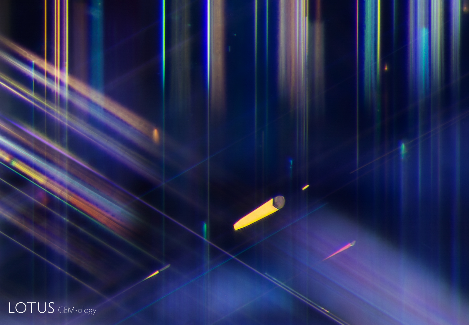
C. A small, rounded crystal accompanied by several elongated, slender tubes stands out in this spessartine garnet. These spindly inclusions evoke a Tim Burton-esque aesthetic.
D. This diagram from Edwin Roedder shows six methods by which primary negative crystals can form. The photomicrographs here are all caused by the capture of a foreign mineral on the surface of a growing crystal (shown in Part F). The impurity creates a shadow behind the impurity, resulting in a tapering cavity behind it in the direction of growth.
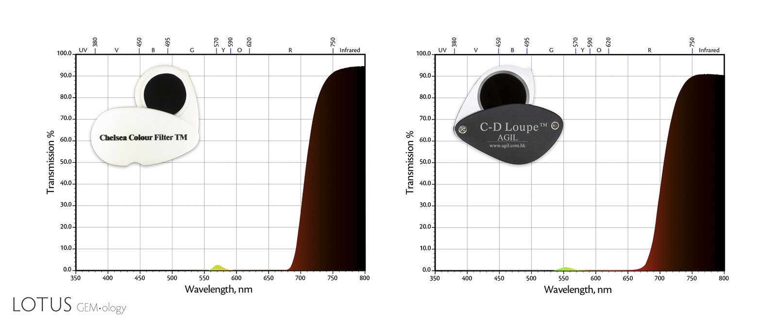
A. The Chelsea filter was developed by B.W. Anderson in 1934 as a method of separating emerald from glass imitations. The advent of synthetic emeralds shortly thereafter made it much less useful, but it still has utility in certain situations (particularly with parcels, which can be quickly scanned). The idea is quite simple, where the filter absorbs all light except narrow bands in the yellow-green (centered at 570 nm) and the red (680–720). Gems that transmit the tail end of the red will appear red, while those that don’t will appear dark. Other filters, such as the AGIL jade filter, are similar.
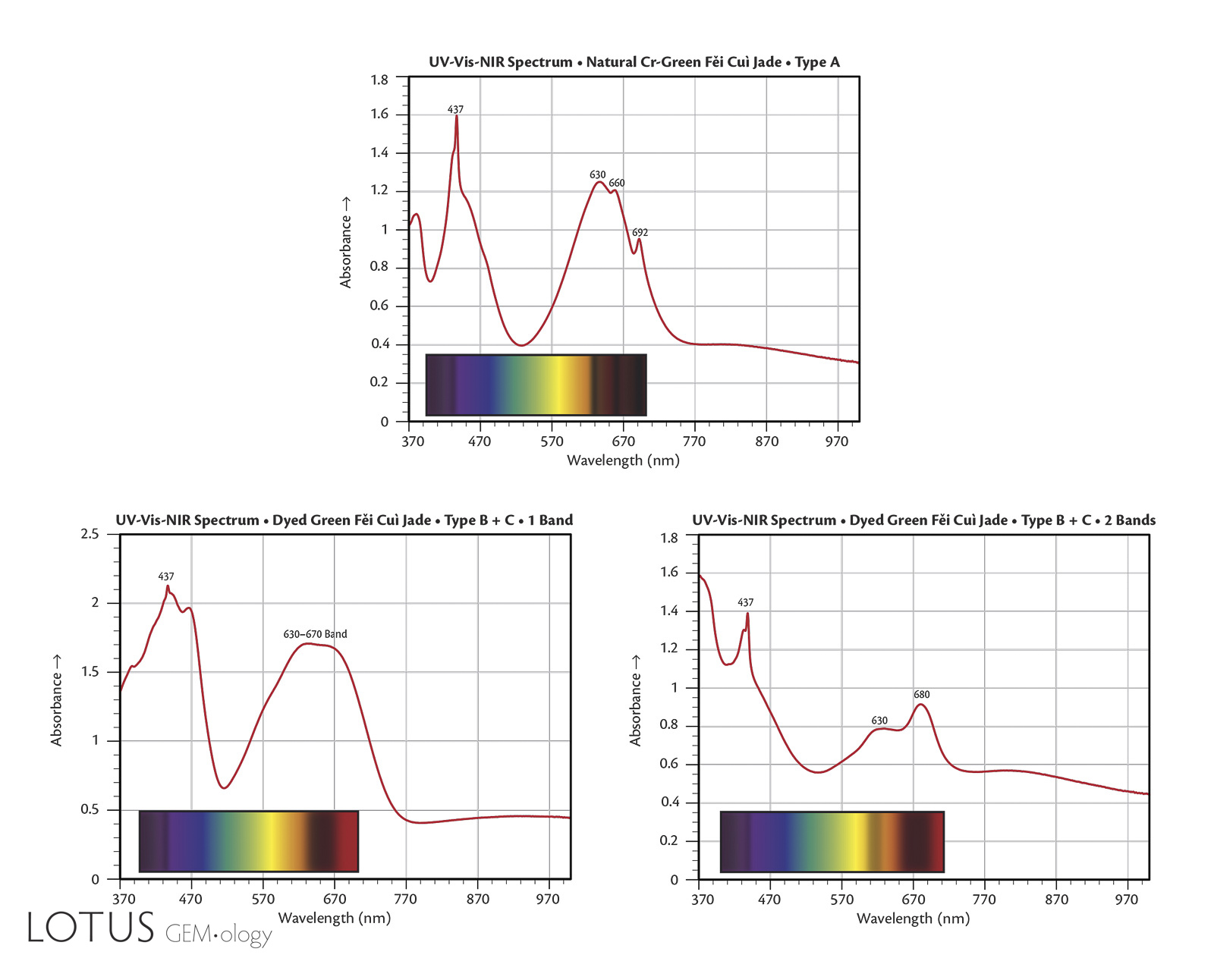
B. The Chelsea filter is of use in identifying some dyed fei cui jade. Natural fei cui that is colored green by chromium completely absorbs the end of the red, thus appearing dark. In contrast, the most common dyes transmit the end of the red, thus appearing red under the Chelsea filter. Unfortunately not all dyes are the same. Thus, if you see red in the Chelsea filter, it suggests the fei cui might be dyed, but if it does not show red, it may still be dyed.
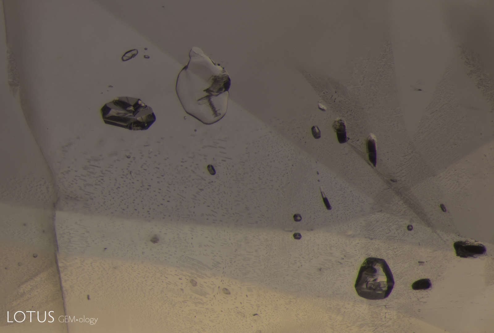
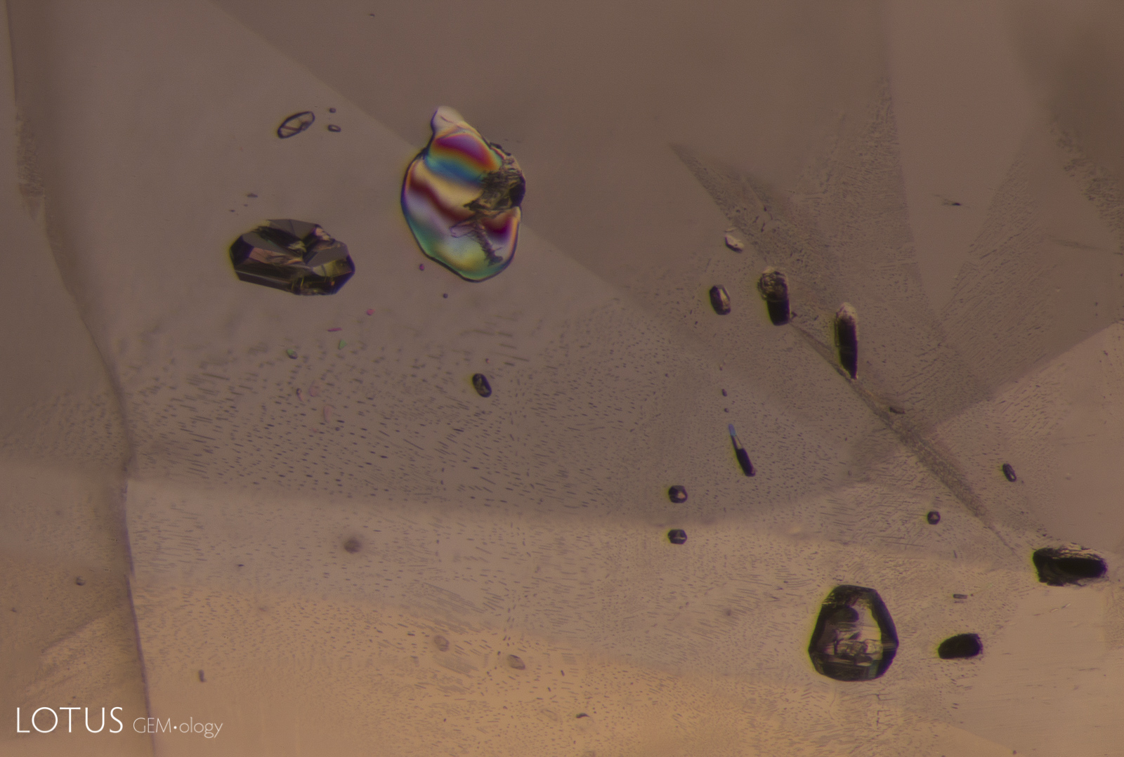
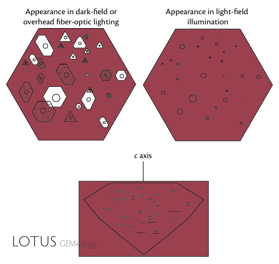
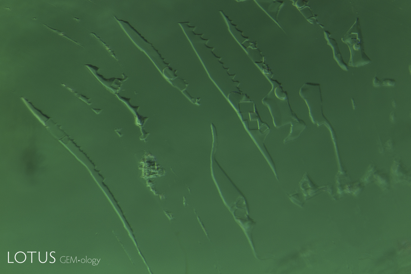
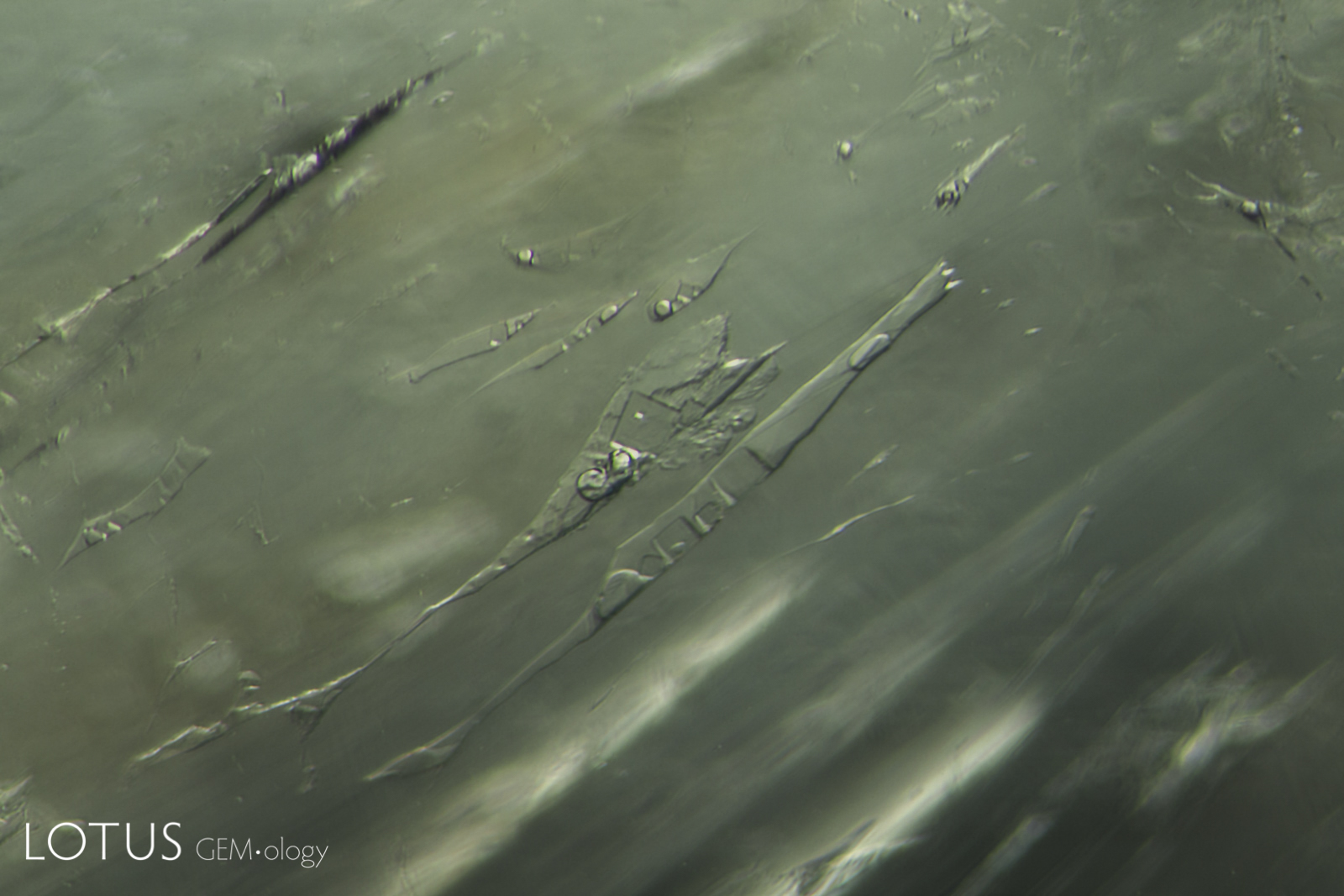
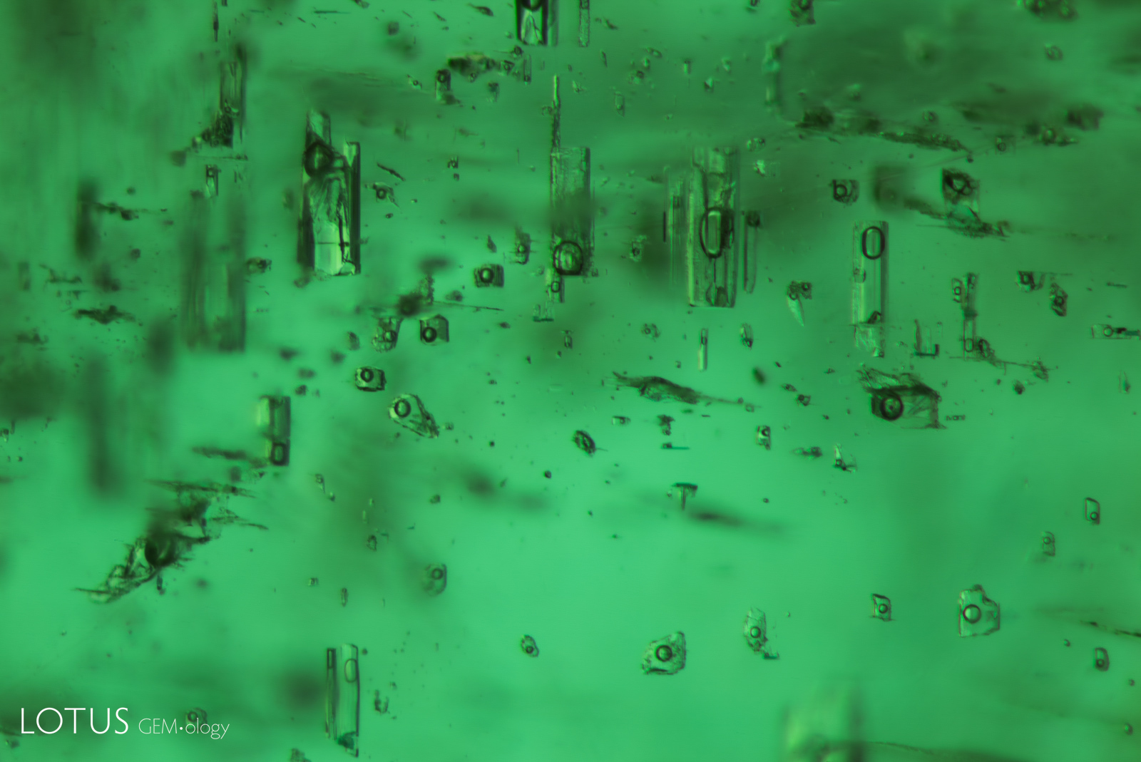
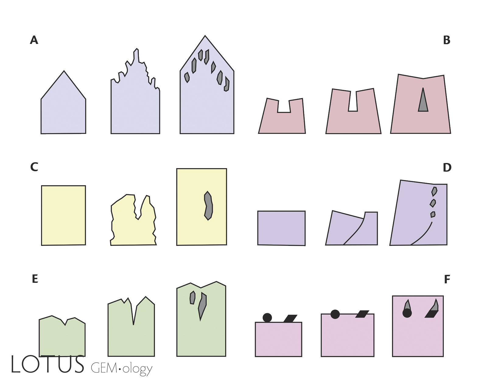
A. Distinguishing solid from negative crystals can sometimes be difficult, but there are a couple things to look for. When viewed in light field illumination, it can be hard to tell a crystal from a negative crystal, as they have a similar appearance.
B. When the same crystals are viewed in crossed polars, if they are doubly refractive, they will display interference colors, clearly separating them from singly refractive and negative crystals.
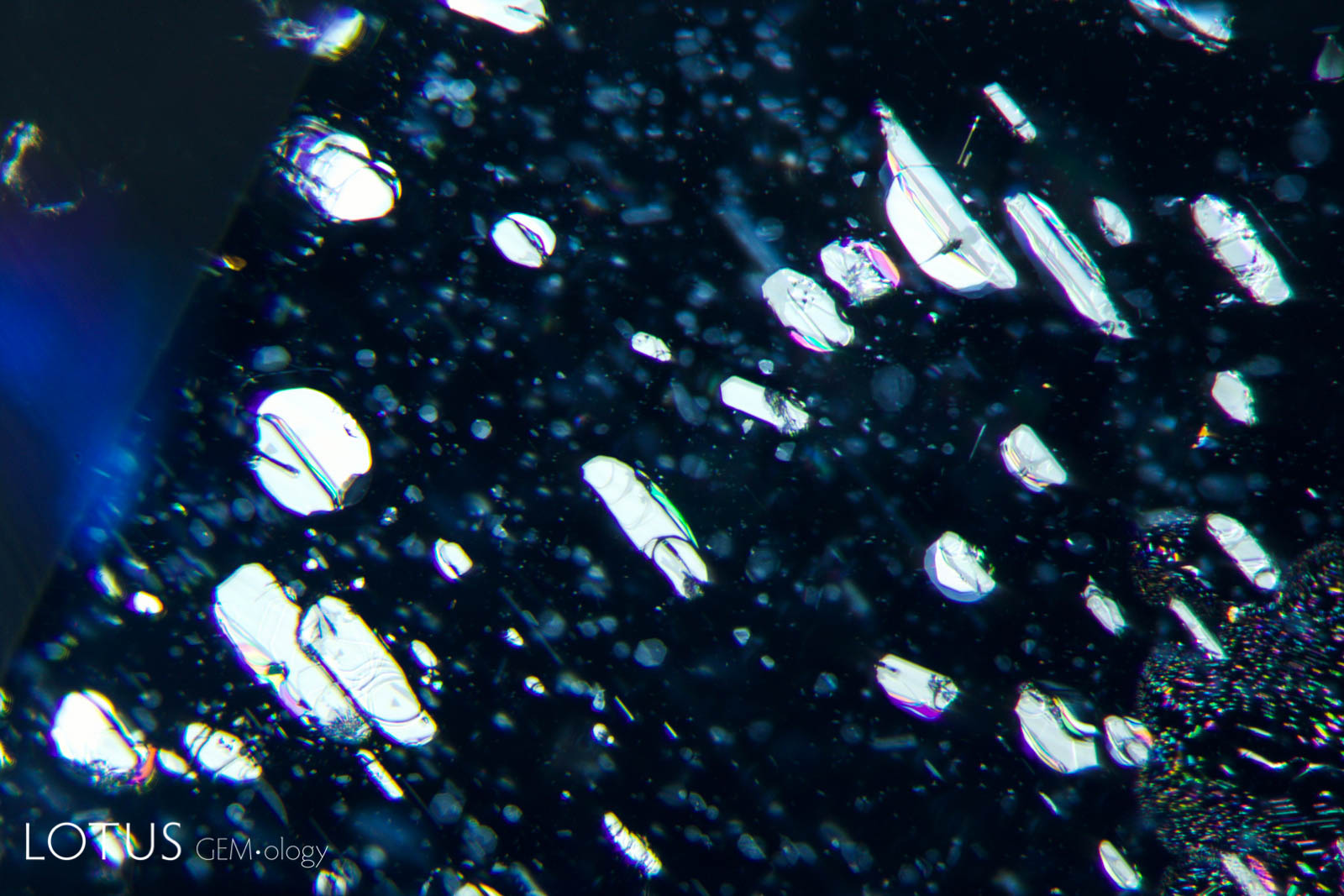
C. Another way to distinguish solid and negative crystals is by their orientation in the host. Solid crystals may be captured at random, but negative crystals always display the faces of their host, because negative crystals are trapped space bounded by the host’s faces. Thus they all have the same orientation. The result is that when light is reflected off their faces, they all reflect at exactly the same angle, as shown in this image.
Note
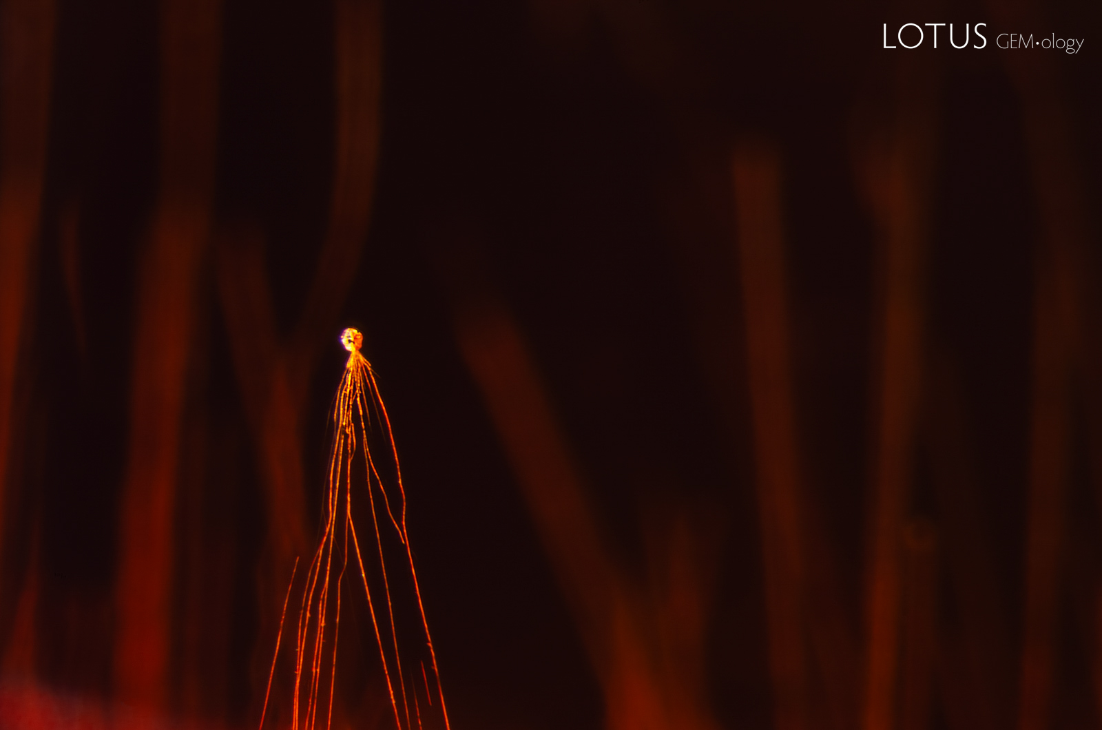
The title graphic was inspired by master photomicrographer John Koivula, who demonstrated this technique of image mirroring to create aesthetic patterns at the International Gemmological Conference in Brazil in 1987.
![]()