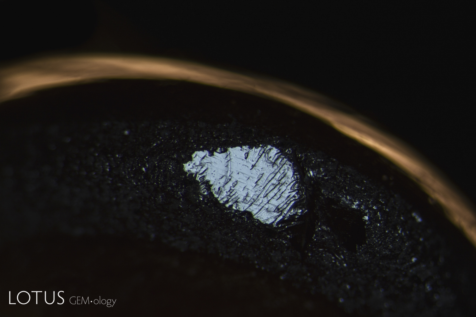
A large black star sapphire was brought in for testing. Two large pits on the base were carefully filled with brown dopping varnish. After removal of the varnish with alcohol, it was found that a large portion of the base had been filled with a lead glass.
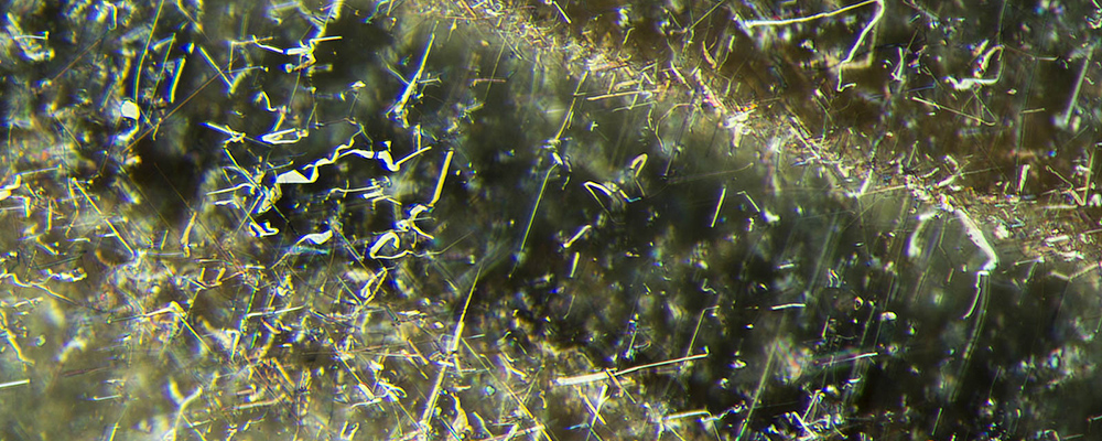
Black star sapphires primarily come from Kenya, Australia and Thailand. Unlike other star sapphires and rubies, the asterism in these stones generally results from exsolution of ilmenite (FeTiO3) and/or hematite (α-Fe2O3) instead of rutile (TiO2). Rutile and ilmenite/hematite will exsolve in different directions, rutile along the second-order hexagonal prism {1120}, with ilmenite along the first-order hexagonal prism {1010}. If both are present in the same stone, a 12-ray star sapphire results (Figure 1).
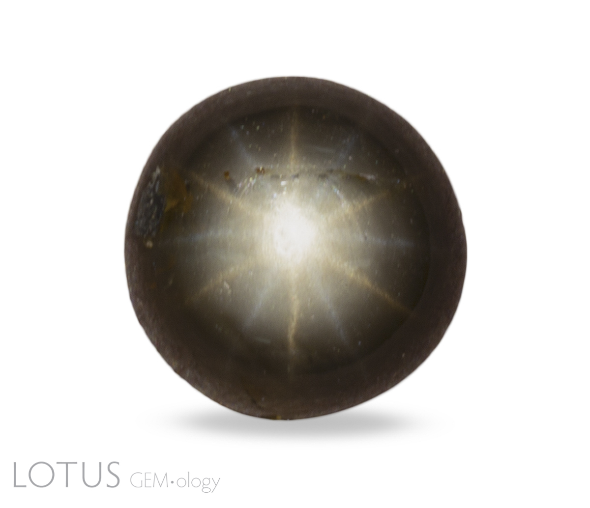 Figure 1. This 12-ray black star sapphire from Chanthaburi, Thailand contains two sets of silk offset 30° from one another. Note that the star created by the rutile is white while the ilmenite/hematite star is slightly yellow. Photo: Chanon Yimkeativong/Lotus Gemology. Click on the photo for a larger image.
The ilmenite/hematite crystals tend to be far more "platy" and thus their layers tend to form planes of weakness. Breakage along those planes is termed parting.
Parting differs from cleavage in that it results from structural planes of weakness due to imperfect growth, rather than inherent weakness. One can think of it like this. If an architect creates plans for a building that results in layers of weakness, we would call that cleavage. But if the plans are sound, but the construction crew did a poor job, that would be parting. Parting results from mistakes in growth; cleavage is a function of design weakness.
 |
 |
| Figure 2. Parting in black star sapphire Left: Basal parting is clearly visible on the bottom of this black star sapphire from Chanthaburi, Thailand. This is caused by the exsolution of platy hematite/ilmenite silk, which produces a lack of adhesion in the basal plane. The two diagonal lines are rhombohedral parting, caused by exsolution of boehmite/diaspore. Field of view: 11 mm Right: Rhombohedral parting on the girdle of a black star sapphire from Chanthaburi, Thailand. Field of view: 4 mm. Photos: Richard W. Hughes; specimens courtesy of Sant Enterprises; |
|
Shellacked
In Thailand, one often sees cavities on the back of black star sapphires filled in with brown dopping shellac (Figure 3). This is because there are often two different parting planes in the stones. One corresponds to the exsolution of ilmenite/hematite in the plane of the basal pinacoid {0001}. The other corresponds to planes of exsolved boehmite/diaspore along the rhombohedron {1011}.
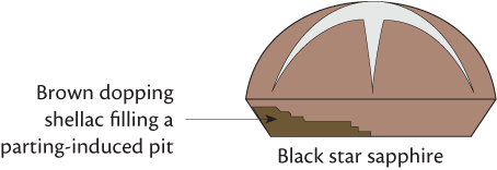 Figure 3. In black star sapphires from Thailand, one often encounters large pits on the back of the stone filled with brown dopping shellac. Illustration: Richard Hughes
In May 2020, a client submitted a large 75-carat black-star sapphire for testing (Figure 4). On the back of the stone were two large pits filled with brown dopping shellac (Figure 5, left). Interestingly enough, the shellac fluoresced bright orange under long wave ultraviolet light (365 nm) (Figure 5, right).
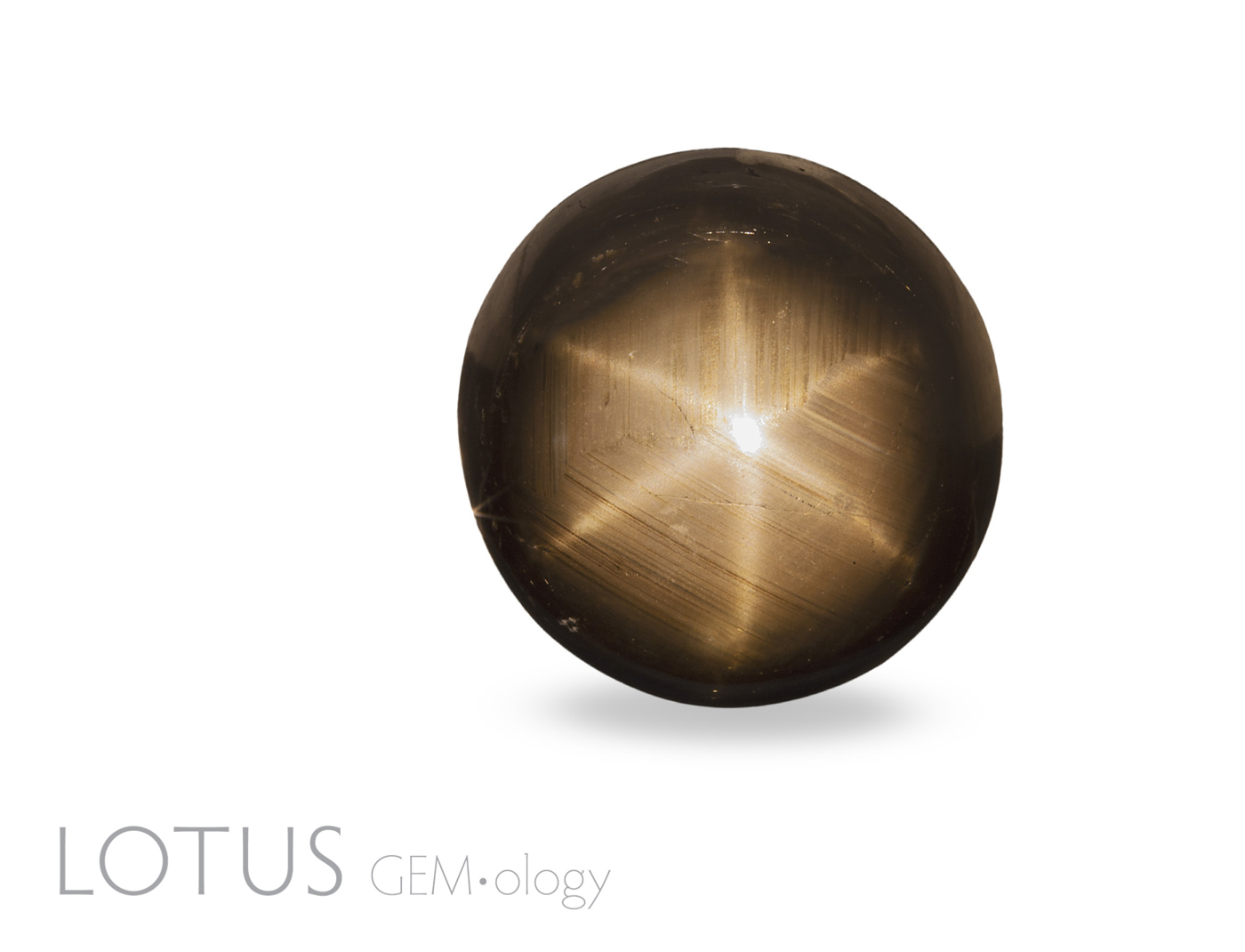 Figure 4. The 75-ct black star sapphire that is the subject of this article. Photo: Wimon Manorotkul
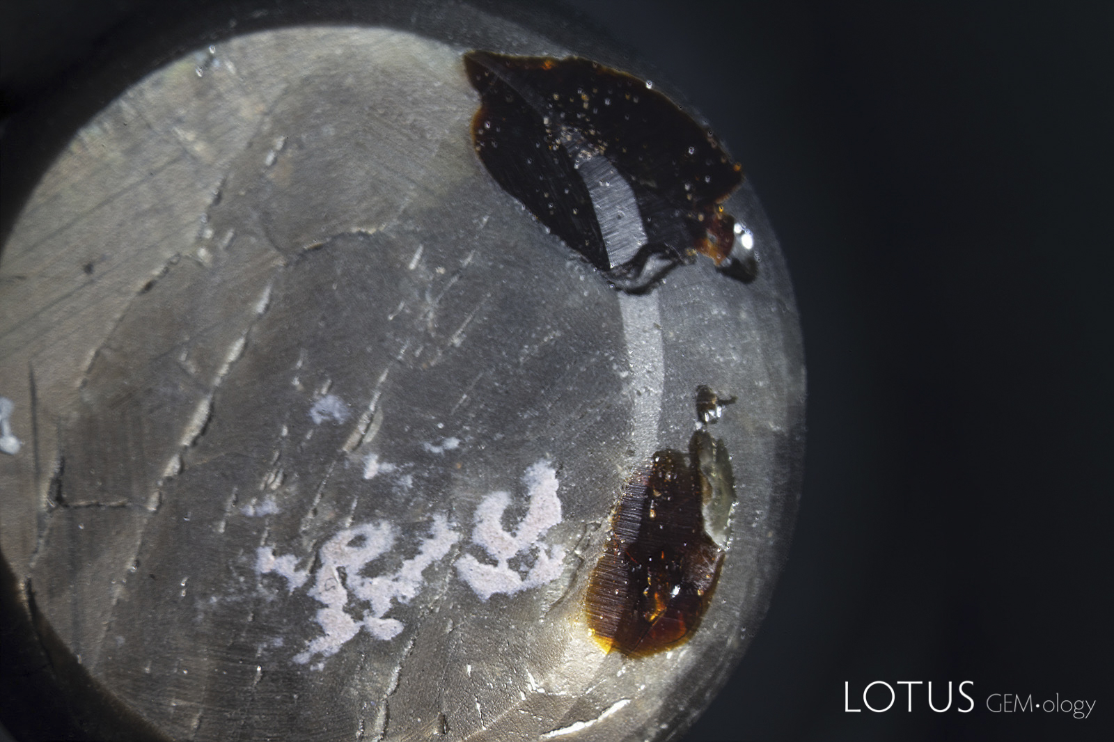 |
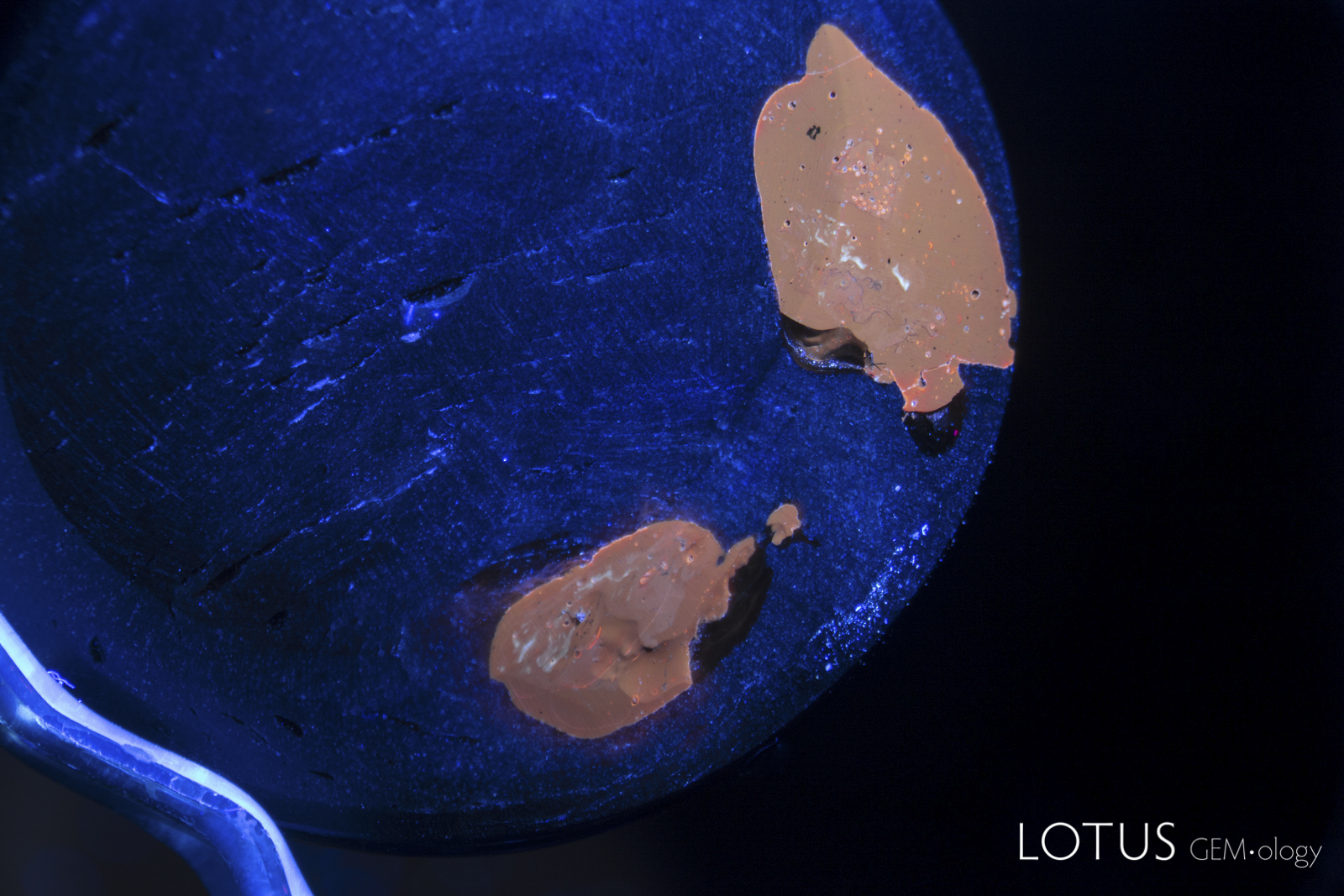 |
| Figure 5. Left: Two large pits on the back of the star sapphire in question. One can see the brown dopping varnish (the white areas are adhesive from tape). Field of view: 30 mm Right: Under long wave ultraviolet light (365 nm), the dopping shellac fluoresced bright orange. Field of view: 27 mm; photos: Richard Hughes; click on a photo for a larger image |
|
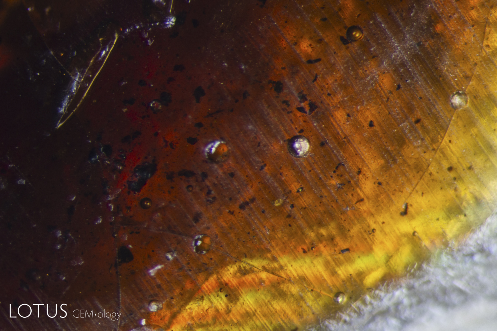 Figure 6. A close-up view of the shellac in the lower pit revealed trapped gas bubbles and black debris. Photo: Richard Hughes; field of view: 5 mm.
We informed the client that we would have to issue the report as a treated stone (Lotus Silver) unless the shellac were removed and offered to remove it with alcohol. Following the client's approval, we removed the shellac.
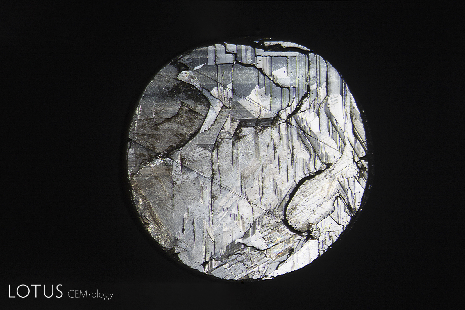
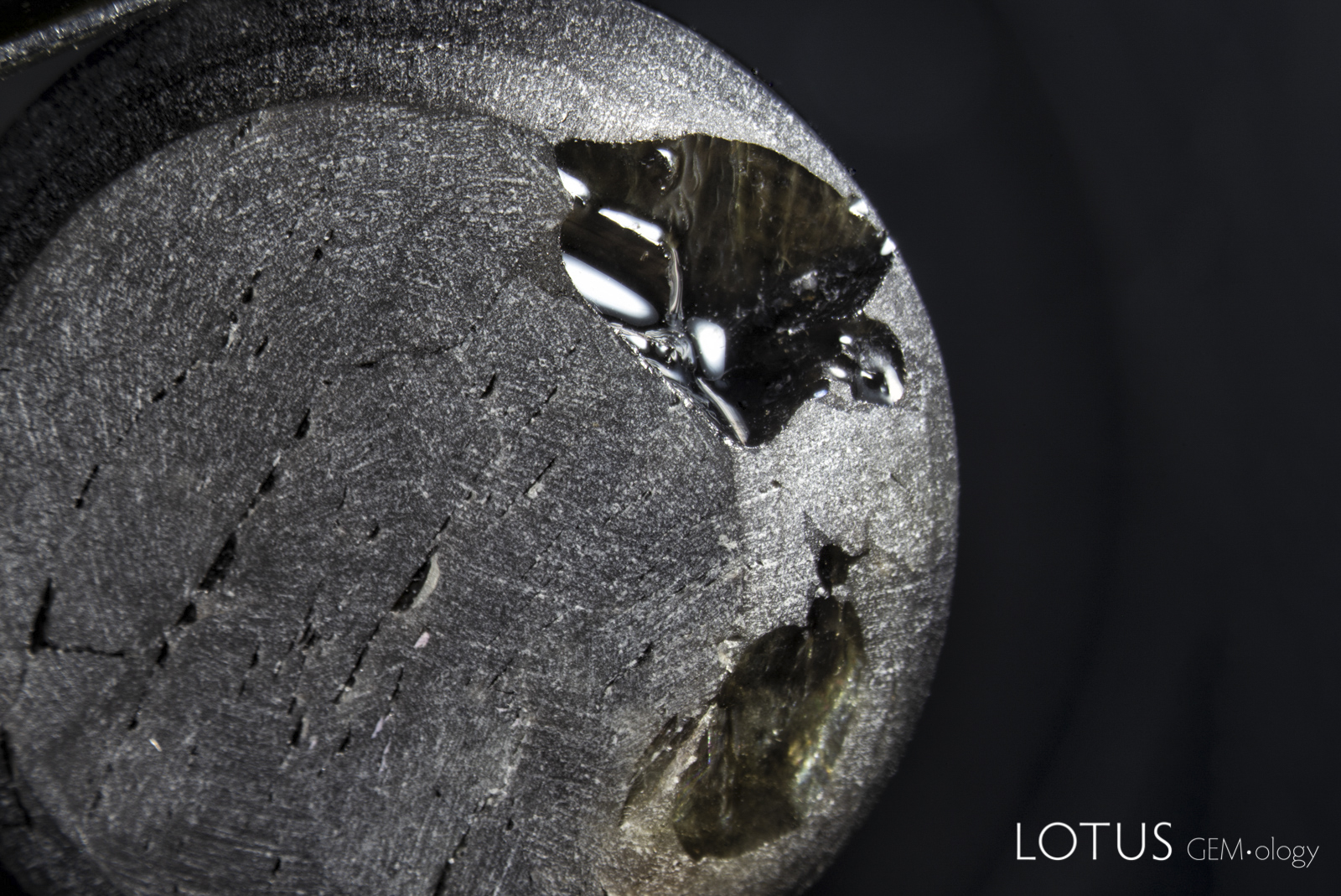 Figure 7. After soaking with alcohol, we were able to completely remove the shellac from the star sapphire. Careful examination of the cavities shows an unusually perfect curved surface, not the type of thing one typically sees on a broken surface of sapphire. Field of view: 30 mm; photo: Richard Hughes.
After removal of the filler, we noticed an unusual curved area exhibiting an unusual flow-like structure that looked suspiciously like glass (Figure 7). Unfortunately, attempting to get a Raman spectrum on the area was unsuccessful. Careful examination showed what looked like gas bubbles trapped between the glassy material and the sapphire below (Figure 8).
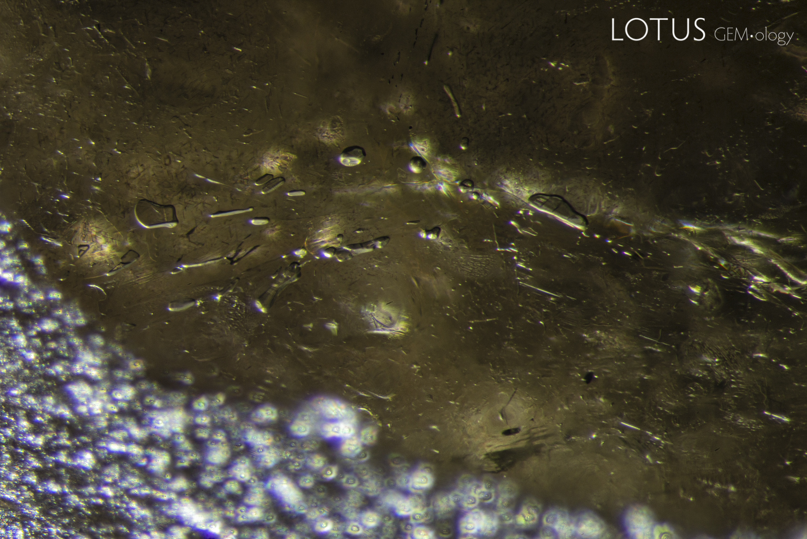 Figure 8. Flattened gas bubbles trapped between the glass filling and the sapphire below. Field of view: 30 mm; photo: E. Billie Hughes.
Under the microscope, we carefully scratched the surface of the cavity with a Number 7 hardness point (Figure 9), which confirmed that the area could not be corundum.
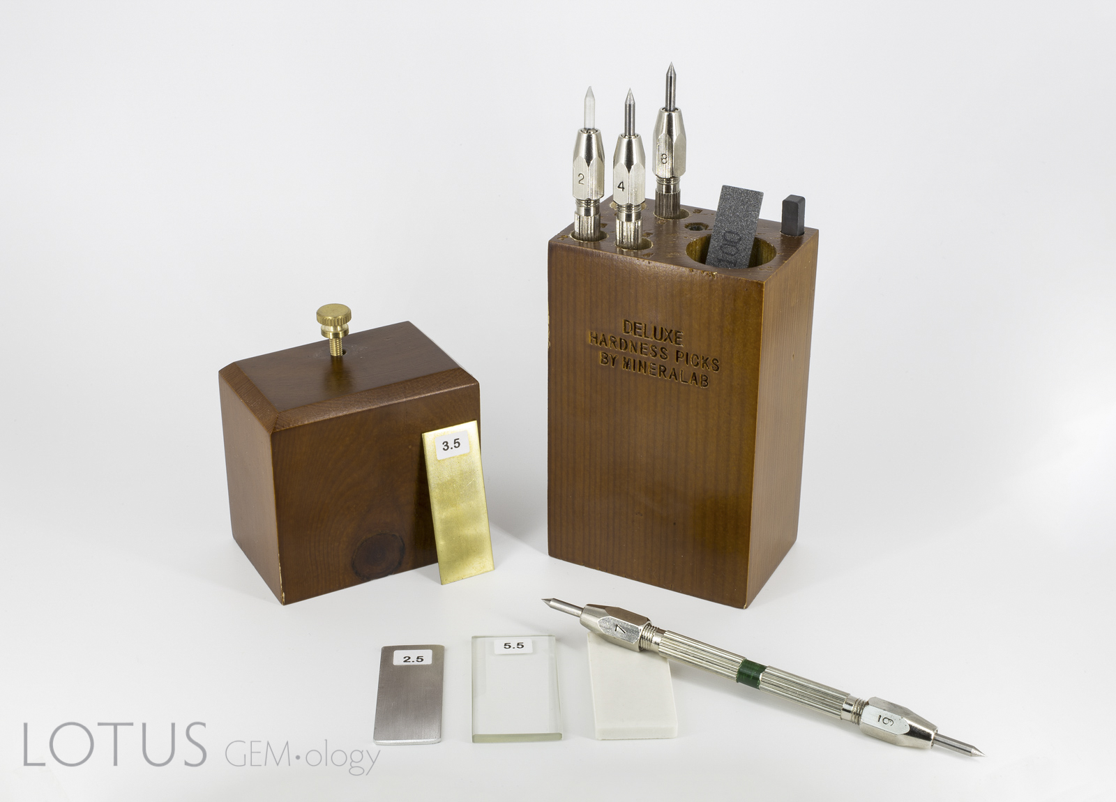 Figure 9. Set of hardness points used by Lotus Gemology. These are easily available for about $110, and include hardness points up to No. 9 on Mohs' Scale. In order to avoid obvious damage, hardness tests should be performed under magnification on inconspicuous locations. Photo: Chanon Yimkeativong/Lotus Gemology.
Following this, we immersed the gem in methylene iodide. This showed the glassy area to cover roughly two-thirds of the base of the cabochon (Figure 10).
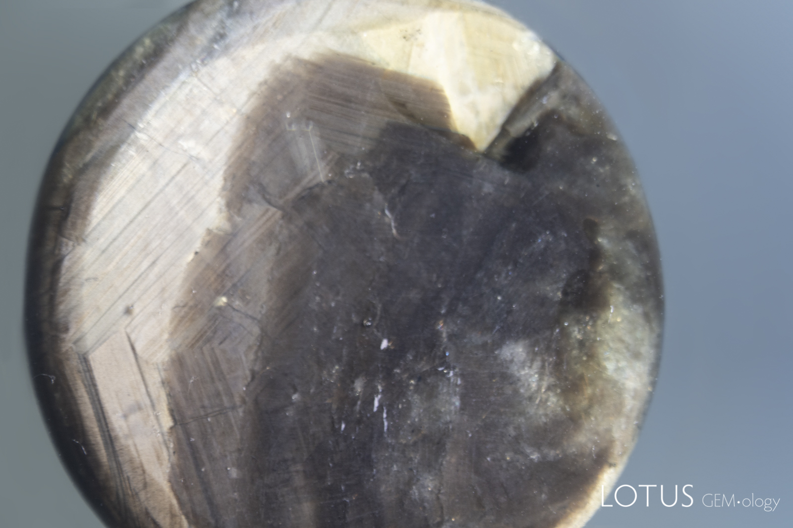 Figure 10. Immersion in methylene diiodomethane (n = 1.741) reveals the extent of the glass filling on the base of the cabochon (the dark area that spreads across a large portion of the base). Field of view: 30 mm; photo: Richard Hughes.
The final test we did was to check the trace element chemistry on the center of the area we suspected to be glass. This revealed a lead (Pb) component of over 3% (Figure 11).
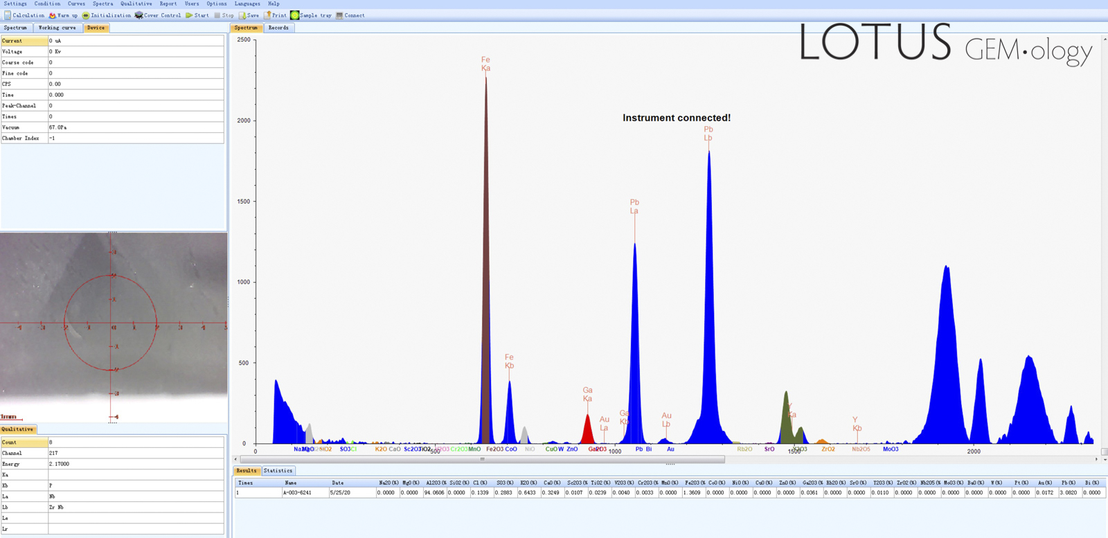 Figure 11. Energy-dispersive x-ray fluorescence (ED-XRF) testing was performed to determine the trace-element chemistry of the center of the sapphire's base. This revealed a lead (Pb) content of about 3%, confirming the filling to be a lead glass.
Conclusion
A variety of tests were brought to bear on this specimen, from the simple (hardness) to the high-tech (ED-XRF). The results of these tests revealed a gem that was treated with two different techniques.
![]()
About the Authors
Richard W. Hughes is one of the world’s foremost experts on ruby and sapphire. The author of many books and over 170 articles, his writings and photographs have appeared in a diverse range of publications, and he has received numerous industry awards. Co-winner of the 2004 Edward J. Gübelin Most Valuable Article Award from Gems & Gemology magazine, the following year he was awarded a Richard T. Liddicoat Journalism Award from the American Gem Society. In 2010, he received the Antonio C. Bonanno Award for Excellence in Gemology from the Accredited Gemologists Association. The Association Française de Gemmologie (AFG) in 2013 named Richard as one of the Fifty most important figures that have shaped the history of gems since antiquity. In 2016, Richard was awarded a visiting professorship at Shanghai's Tongji University. 2017 saw the publication of Richard and his wife and daughter's Ruby & Sapphire • A Gemologist's Guide, arguably the most complete book ever published on a single gem species and the culmination of four decades of work in gemology. In 2018, Richard was named Photographer of the Year by the Gem-A, recognizing his photo of a jade-trading market in China, while in 2020, he was elected to the board of directors of the Accredited Gemologists Association and was appointed to the editorial review board of Gems & Gemology and The Australian Gemmologist magazine. In 2022, Richard published Jade • A Gemologist's Guide, while 2024 brought Broken Bangle • The Blunder-Besmirched History of Jade Nomenclature. His jade trilogy was completed in 2025 with his translation of Heinrich Fischer's Nephrite and Jadeite.
E. Billie Hughes is Co-Founder and Managing Director of Lotus Gemology. She oversees the company's day-to-day operations while continuing gemological research and laboratory work. After graduating from UCLA in 2011, Billie became a Fellow of the Gemmological Association of Great Britain (FGA) in 2013. Her research focuses on ruby and sapphire, including low-temperature heat treatment, and she has authored and co-authored articles in leading gemological journals. An accomplished field gemologist, she has traveled to gem deposits around the world, including nearly every major ruby and sapphire locality.
Billie is an internationally recognized educator who has lectured for trade organizations, museums, and luxury jewelry houses. She has collaborated extensively with Van Cleef & Arpels on educational programs and lectures. An award-winning photographer and photomicrographer, her images have received honors in the Nikon Small World and Gem-A competitions and have appeared in publications including National Geographic and Forbes. She is also the creator of Hyperion, Lotus Gemology's online inclusion database, reflecting her commitment to making gemological knowledge more accessible.
Billie developed an interest in gemstones from an early age, accompanying her parents on expeditions to mines and gem-producing regions around the world. That lifelong passion for fieldwork, laboratory research, education, and photography continues to shape her work at Lotus Gemology today.
Wimon Manorotkul has been involved with gems and gemology since 1979, as a lab gemologist, instructor and photographer. She is an Accredited Gemologist from Bangkok's Asian Institute of Gemological Sciences and for many years directed their lab. Wimon also qualified as a Fellow (with honors) of the Gemmological Association of Great Britain. A skilled gem photographer, her images have been featured in books and magazines around the world, particularly Ruby & Sapphire: A Collector's Guide, along with Ruby & Sapphire: A Gemologist's Guide and Inside Out | GEM• ology Through Lotus-Colored Glasses. Wimon not only photographs gems, jewelry and mineral specimens, but is also an expert photomicrographer. In 2013, she founded Lotus Gemology with her husband, Richard Hughes, and daughter, E. Billie Hughes.
Notes
Written in May–June 2020. An abbreviated version was published in Gems & Gemology (Fall 2020, pp. 442–443) (Fall 2020, pp. 442–443).
References
- Bui, T.N. (2018) Peculiar trapiche-like pattern in a gold-sheen sapphire. Journal of Gemmology, Vol. 36, No. 4, pp. 289–291; RWHL.
- Bui, T.N., Deliousi, K., Corte, K., de and Gauthier, J.-P. (2016) Star sapphire showing a variable number of rays. Journal of Gemmology, Vol. 35, No. 3, July, pp. 197–199; RWHL.
- Bui, T.N., Deliousi, K., Malik, T.K. and Corte, K., de (2015) From exsolution to 'gold sheen': A new variety of corundum. Journal of Gemmology, Vol. 34, No. 8, October, pp. 678–691; RWHL*.
- Moon, A.R. and Phillips, M.R. (1984) An electron microscope study of exsolved phases in natural black Australian sapphire. Micron and Microscopica Acta, Vol. 15, No. 3, pp. 143–146; RWHL.
- White, J.S. (1979) Boehmite exsolution in corundum. American Mineralogist, Vol. 64, Nos. 11–12, pp. 1300–1302; RWHL*.
![]()