A look into the world of spinel inclusions that goes beyond simple octahedral crystals.
Spinel Inclusions
When gemology students are taught about spinel, one of the first things they are told to look for are octahedral crystals. These echo the form of spinel crystals and look like little bipyramid shapes inside the spinel. Although they are a classic, diagnostic feature, there are many other interesting inclusions to be found in the spinel realm. The following are a few examples the author has had the opportunity to photograph in the laboratory.
 Above is an octahedral crystal, a typical feature to look for when identifying spinel. Darkfield + oblique fiber optic illumination. Photo: E. Billie Hughes
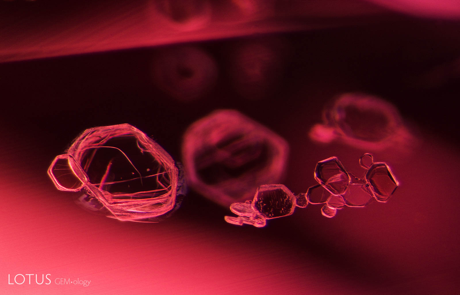
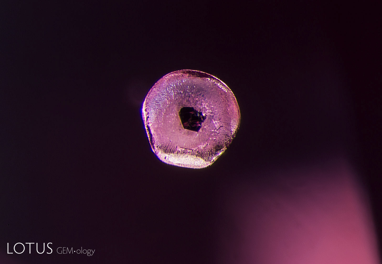 We are sure Homer Simpson would love this one! This donut-shaped inclusion is an apatite crystal, common in spinel from both Burma and Sri Lanka. The black flake in the center is actually a graphite crystal, creating what master photomicrographer John Koivula has whimsically nicknamed “belly button” apatite crystals. Oblique fiber optic illumination. Photo: E. Billie Hughes
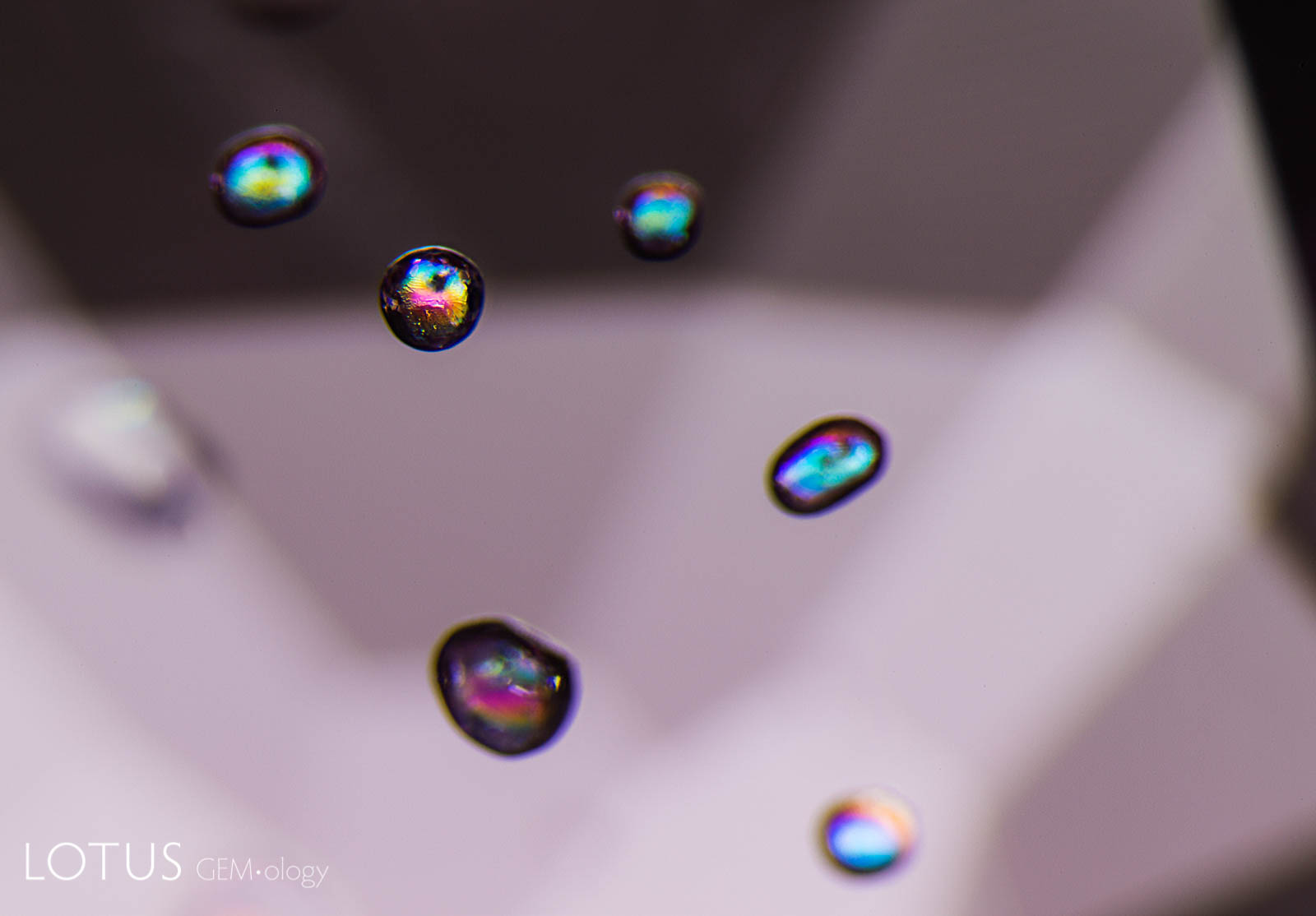 More apatite crystals on display. When viewed under crossed polars as shown above, the crystals display a rainbow of interference colors. Photo: E. Billie Hughes
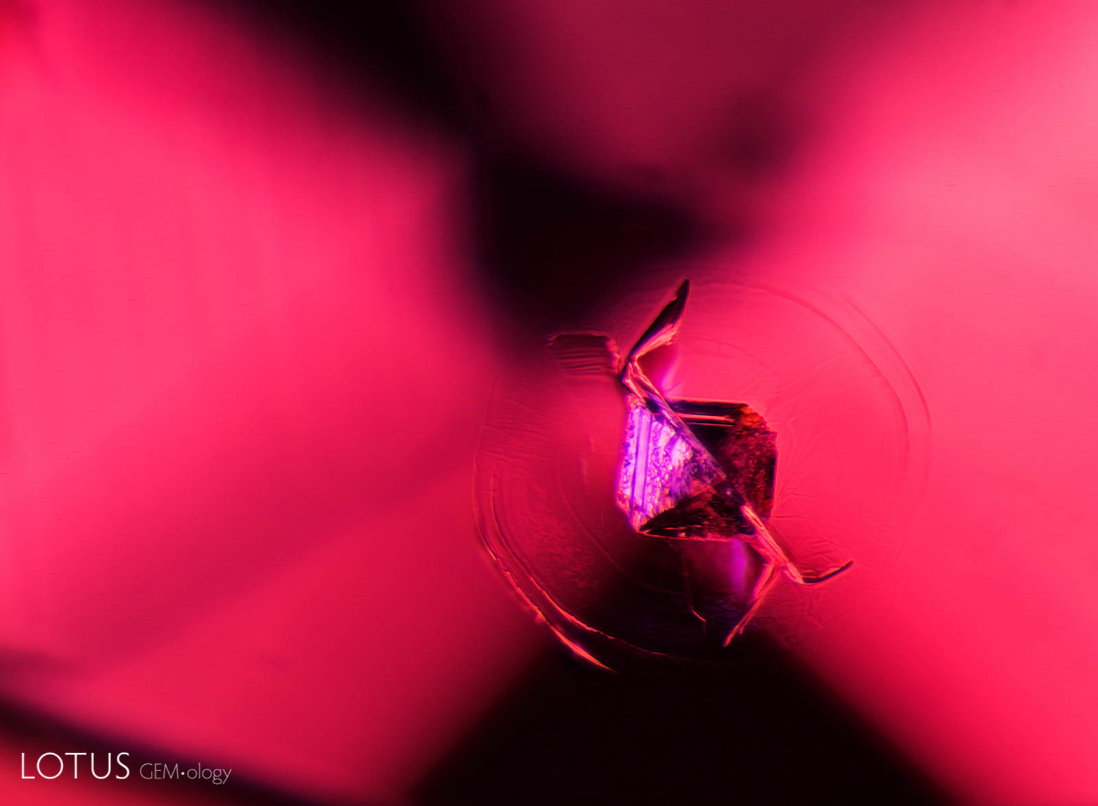
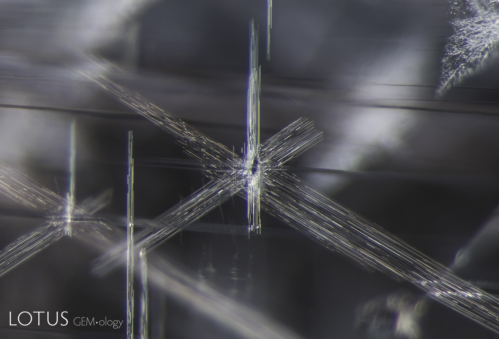 Stellate dislocations decorate the interior of this spinel from Vietnam. We also often see such dislocation needles in material from Sri Lanka. Diffuse oblique fiber optic illumination. Photo: E. Billie Hughes
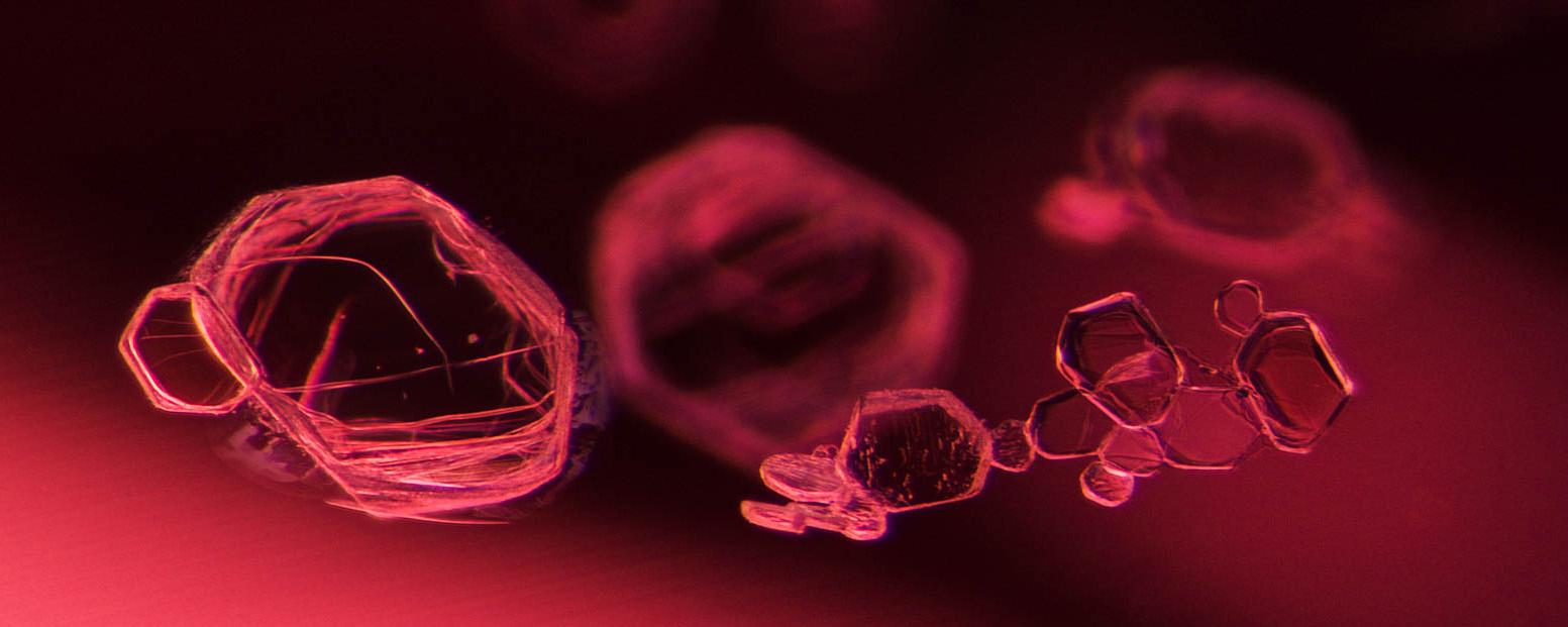
 Small transparent crystals form clusters in a Vietnamese spinel. These tabular crystals are transparent and doubly refractive. Dark field illumination. Photo: E. Billie Hughes
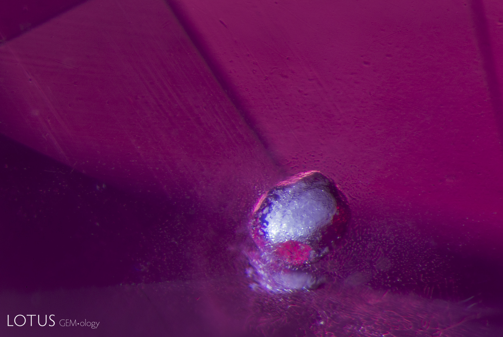
 At first glance the tiny exsolved particles in this spinel from Mahenge, Tanzania, may look like specks of dust. Upon close observation we can see that they are actually scattered throughout the stone, not on the surface. These dust-like particles are a common feature in Mahenge spinel. Diffuse oblique fiber optic illumination. Photo: E. Billie Hughes
 Another common feature in spinel from Mahenge, Tanzania, are this fine needle-like inclusions. These are most easily seen with fiber optic illumination, as shown here. Diffuse fiber optic illumination. Photo: E. Billie Hughes
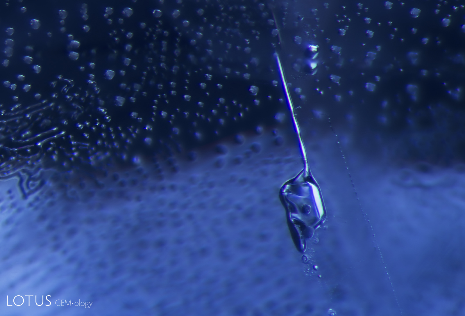
 This melted crystal shows a “frosty” appearance similar to that of a snowball. The vast majority of spinels that we see in our lab are untreated, but this is a rare example of a heated spinel. The host of this melted crystal is a Mahenge, Tanzania, spinel. Darkfield + Diffuse oblique fiber optic illumination. Photo: E. Billie Hughes
 In another example of a heated spinel, this time in a cobalt-diffused stone, a melted crystal stands out against a backdrop of tiny heat-altered octahedra. Note how the faces of the crystals all display a highly reflective, glassy appearance, and the edges are rounded rather than angular. Dark-field illumination. Photo: E. Billie Hughes
![]()
About the author
E. Billie Hughes is Co-Founder and Managing Director of Lotus Gemology. She oversees the company's day-to-day operations while continuing gemological research and laboratory work. After graduating from UCLA in 2011, Billie became a Fellow of the Gemmological Association of Great Britain (FGA) in 2013. Her research focuses on ruby and sapphire, including low-temperature heat treatment, and she has authored and co-authored articles in leading gemological journals. An accomplished field gemologist, she has traveled to gem deposits around the world, including nearly every major ruby and sapphire locality.
Billie is an internationally recognized educator who has lectured for trade organizations, museums, and luxury jewelry houses. She has collaborated extensively with Van Cleef & Arpels on educational programs and lectures. An award-winning photographer and photomicrographer, her images have received honors in the Nikon Small World and Gem-A competitions and have appeared in publications including National Geographic and Forbes. She is also the creator of Hyperion, Lotus Gemology's online inclusion database, reflecting her commitment to making gemological knowledge more accessible.
Billie developed an interest in gemstones from an early age, accompanying her parents on expeditions to mines and gem-producing regions around the world. That lifelong passion for fieldwork, laboratory research, education, and photography continues to shape her work at Lotus Gemology today.
Notes
First published in the Journal of The Gemmological Association of Hong Kong, 2017, Vol. XXXVIII, pp. 41–44).
References & further reading
- Gübelin, E.J. and Koivula, J.I. (1986) Photoatlas of Inclusions in Gemstones. Zurich, Switzerland, ABC Edition, 532 pp.
- Gübelin, E.J. and Koivula, J.I. (2005) Photoatlas of Inclusions in Gemstones, Volume 2. Basel, Switzerland, Opinio Publishers, 830 pp.
- Saeseaw, S., Wang, W. et al. (2009) Distinguishing Heated Spinels from Unheated Natural Spinels and from Synthetic Spinels. Online report, April 2, Gemological Institute of America, 13 pp.
- Schmetzer, K., Gübelin, E. et al. (2000) Oriented inclusions in spinels from Madagascar. Journal of Gemmology, Vol. 27, No. 4, pp. 229–232
![]()