This article traces the development of the modern gemological laboratory from 1925 to the present date, a 100-year journey that has seen not just a gradual and then explosive evolution in the application and maturity of the science, but importantly the standing that gemological laboratories have enjoyed within the gemstone industry and the scientific community. The passionate people involved have made tremendous contributions to the understanding of gem materials and their willingness to embrace the latest technology as well as attract successive generations to the field has made the study of gem materials within gemological laboratories one of the most exciting theatres of life.
In 1973 this author attended a presentation in London’s Goldsmiths Hall given by B.W. Anderson. The title of the presentation was 1925… and all that (Anderson, 1973). Anderson recounted that:
The forty-six years during which I oversaw the Laboratory of the London Chamber of Commerce can be considered conveniently to consist of four periods. The first (1925–1930) was concerned almost exclusively with pearl-testing. In the second period, ushered in by the World Trade Depression, we (I had been joined by C.J. Payne in 1928) extended and improved our techniques for gem-testing, and used the leisure forced on us by the Depression to carry out some fruitful research. This was ended by the outbreak of war, and the war, of course, was a very distinctive period of its own. And the last period began in 1946, when, faced with an enormous increase in work, Robert Webster and Alec Farn were added to strengthen our well-balanced team of four thereafter working together for the next twenty-five years.
 |
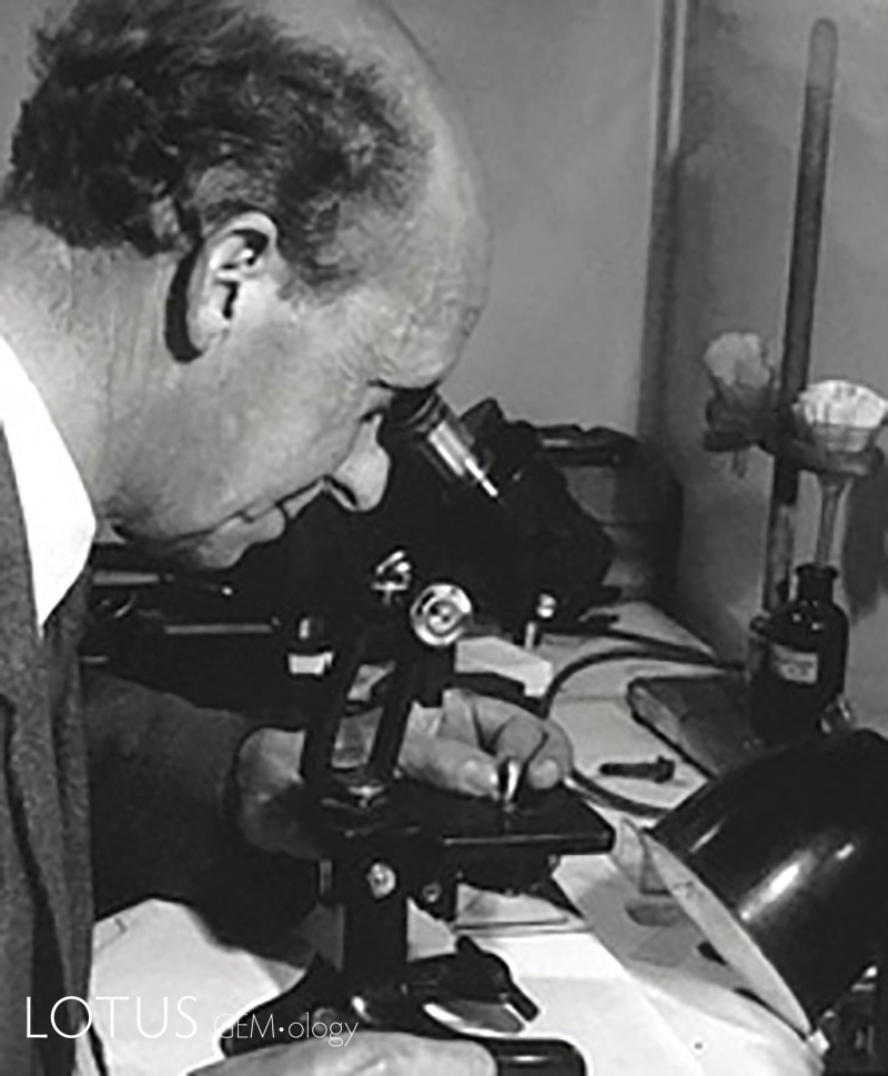 |
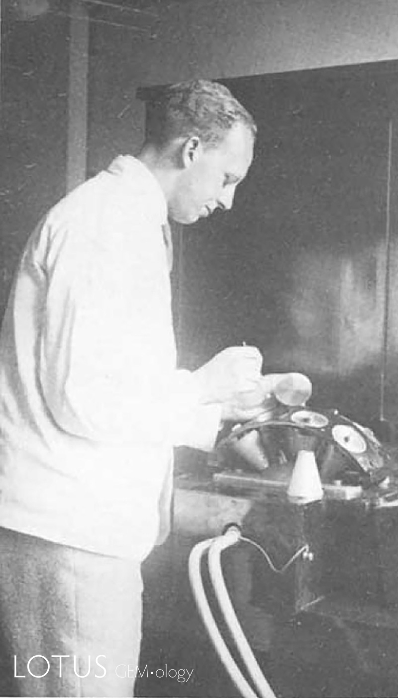
| Figure 1 (left). B.W. Anderson pictured setting up pearls on the Laue diffraction cones dated in the late 1920’s. Figure 2 (right). B.W. Anderson using a Beck Greenough stereo binocular microscope in the 1970’s. |
|
A Century of Work
It has been 100 years now since Anderson was given the task of establishing the world’s first specialised gemological laboratory where the initial focus was the establishment of test methods that separated the then new bead cultured pearl from natural pearls. Anderson and the London Laboratory were joined in the race to stabilise the pearl markets in 1929 by a new laboratory in Paris, under the guidance pf Georges Gobel, the Service Public du Contrôle des Diamants, Perles Fines et Pierres Précieuses de la Chambre de Commerce et d’Industrie de ParisI (Anderson, 1981; Scarratt & Karampelas, 2020). Both laboratories initially focused their efforts on the use of Simon & René Bloch’s pearl endoscope (Poirot & Gonthier, 1998; Webster, 1957) that was manufactured in Paris, and X-ray Laue diffraction (Figure 1) (Webster, 1955), where a combination of the two methods established that bead-cultured pearls could be separated from natural pearls. This was added to by the development by GIA in the 1940’s of the “pearloscope" (Zhou, 2021), an instrument that combined microscopy, the endoscope and the observation of candling[1]. In later years with the introduction of non-bead cultured pearls X-ray microradiography was introduced (Alexander & Sherwood, 1940; Alexander, 1941a–b). As the period progressed gemology became a popular pastime, and sometimes even an obsession in Europe the USA and elsewhere with excellent papers being published by B.W., Anderson and C.J., Payne and such great names and Alfred and W.F. Eppler, E. Gübelin, H. Tillander, O. Dragsted, K. Schlossmacher, A. Chikayama, R.M. Shipley, R. Crowningshield and R.T. Liddicoat who were prolific authors in trade and mineralogical publications that garnered and spread gemological knowledge across the world (Anderson, 1931, 1935a–b, 1937a–b; Anderson & Payne 1937a–b, 1939a–b,; Eppler ,1928, 1933, 1935, 1938; A. Eppler 1934; Gübelin, 1939, 1940a–c; Liddicoat & Ball 1941; Shipley & Liddicoat 1941).
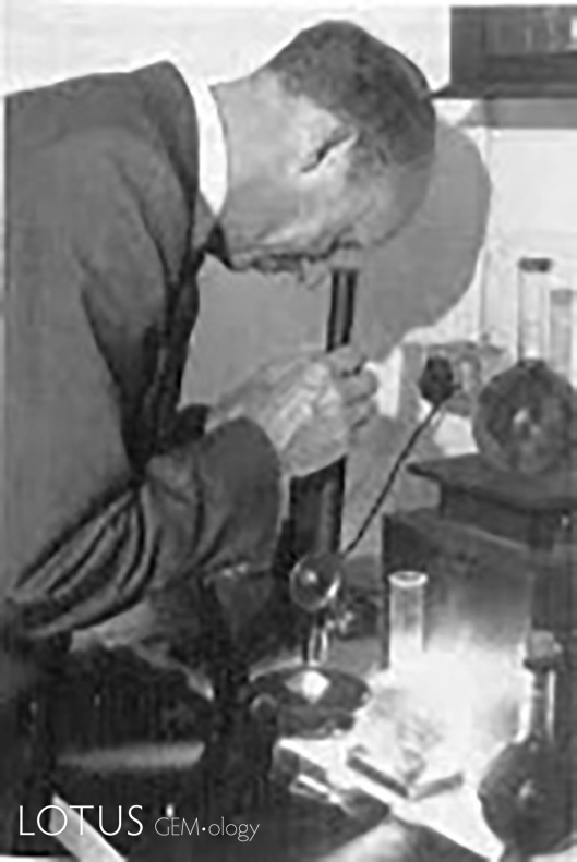 Figure 3. Anderson using the hand-held spectroscope, in the 1970’s.
After 1930 came the global “Great Depression” and between then and the beginnings of World War II, with business being at a low ebb Anderson and Payne who were both academically oriented, Anderson the chemist and Payne the mathematician, used their time to establish the correct refractive indices and SG’s of gem materials that had been erroneously published in earlier gemological and industry texts, greatly assisted with the publication of Larsen’s Microscopic Determination of Nonopaque Materials (Larsen, 1921). In doing this there was a realisation that gemologists needed further a development of the refractometer devised by G.F Herbert-Smith (Smith, 1913) and certainly one less complex in use than the precise Abbe Pullfrich unit that they had been using to determine RI’s often to five places of decimal. This resulted in the development of new refractometers with various kinds of glass and mineral hemispheres or prisms including the Anderson-Payne spinel refractometer and a model with a diamond prism (Anderson et al., 1940; Burbage & Anderson, 1942) the development of the 1.81 refractive index liquid by Anderson and the study of heavy liquids for SG determinations (Mitchell, 1980). The 1940’s and 50’s also heralded the introduction of the hand-held spectroscope (Anderson, 1944a–b, 1950, 1970, 1972; Anderson & Payne, 1954–1956, 1998) a gemological tool that completed along with the long working distance
Beck Greenough binocular stereo microscope (Figure 2) the requirements for a gemological laboratory. Anderson developed a unique method of laboratory use for the spectroscope by employing a 500-watt projection bulb the light from which was cooled and focused by a water (or copper sulphate solution if a filter was needed) filled glass flask onto the mirror of a monocular microscope. Light reflected from the mirror passed up through the microscope stage where the stone under examination was held. The light, having passed though the stone, being examined by the spectroscope in the position of the ocular – which had been remove (Figure 3).
Enter the Synthetics
While synthetic[2] ruby had been commercially available since the late 1800’s synthetic versions of gems were not deemed a great threat to the market until it was realized in the 1950’s and 60’s that significant quantities of synthetic ruby melee had gone unnoticed in art nouveau and art deco line bracelets and various other items of jewellery for several decades. During this period vast quantities of Burmese ruby melee that needed to be tested were added to the workloads of laboratories and these required new examination techniques. Fortunately the synthetic rubies involved were of the Verneuil type, where separation required only simple microscopic observations with the melee lined up on a glass slide in partial immersion in a liquid such as benzyl benzoate[3]. However, it was not long before flux grown synthetic rubies and emeralds added to industry problems and further innovative instrumentation and techniques were needed.
While other microscopes in differing configurations were developed through the 60’s and 70’s, the GIA’s Gemolite proved the be the most popular for diamond and colored stone examinations and assisted greatly with identifying the growing list of synthetic gem materials, at this time including, ruby, sapphire, emerald, alexandrite, and opal.
However, the use of immersion liquids to better understand growth phenomena was not easy with vertical oriented microscopes such as the Gemolite and this inspired the design of horizontal gemological microscopes (Kiefert & Schmetzer, 1991a–c; Nelson 1985) and the use of the same with different optics to project growth phenomena images onto a screen for education purposes (Nelson, 1991). When optical fibre lighting was added to for use with the microscope (Koivula, 1982)as well as with the spectroscope, in the late 70’s the basic tools of most gemological laboratories, which were now proliferating globally, were set. The use of the various refractometers, hand-held spectroscopes, dichroscopes, gemological microscopes and optical fibre lighting were gemological mainstays throughout the 60’s and 70’s. However, with the trade realising the capabilities of gemological laboratories, or lack thereof, pressure was added to expand their scope of work.
As synthetic emeralds entered the market filters such as the Chelsea Filter and others (Anonymous, 2024; Liddicoat, 1984) became the first line of defence for most gemologists and the fledgeling laboratories that were growing globally as a result of the increased availability of gemological education.
In the early 1970’s laboratories began to look towards solving gemological issues that were not solvable with the standard instrumentation and in particular opaque gems such as the jade-like minerals, by installing and employing Debye-Scherrer X-ray Powder Diffraction (XRD) cameras (Koivula & Fryer, 1984; Lind et al., 1983; Nelson, 1960; Scarratt, 1987c).
During the late 50’s and 60’s Robert Crowningshield in New York, and others, were increasingly faced with the appearance of diamonds treated by irradiation followed by annealing (Crowningshield, 1957, 1969). It was discovered then that absorption lines seen in the visible spectra of diamonds became sharper if the diamond was cooled. Wishing to understand and develop this observation stories abounded at the time of Anderson walking to Smithfield’s meat market[4] where he filled his briefcase with dry ice to cool down diamonds for spectroscopic observations – strangers in the street were in wonderment of his briefcase that became increasingly frosted as he walked back to the laboratory in his typical three piece pin-striped suite. In the early 70’s Alan Collins and the team from the Wheatstone Physics (now Innovation) laboratory at King College London became involved with diamond research and proposed that the London laboratory use liquid nitrogen gas to cool diamonds under examination. This led to the construction of an optical bench and cooling system whereby diamonds could be held at -160 °C for extended periods during the hand-held spectroscopic examination (Scarratt, 1979). Shortly after this George Bosshart of the newly formed SSEF laboratory devised a. evacuated glass vessel with parallel quartz windows that allowed the temperatures to be stably reduced to that of liquid nitrogen but now using a Pye Unicam UV visible spectrophotometer (Bosshart, 1989). This, in the early 1980’s also heralded the use of UV visible spectrophotometry in both the GIA and London Laboratories, not just for diamonds (Collins, 1982; Scarratt, 1984, 1986b; Woods & Collins, 1986) but also for colored stones, where Bosshart again led the field with his observations on UV transparency differences, at the time, between natural and synthetic rubies (Bosshart, 1982).
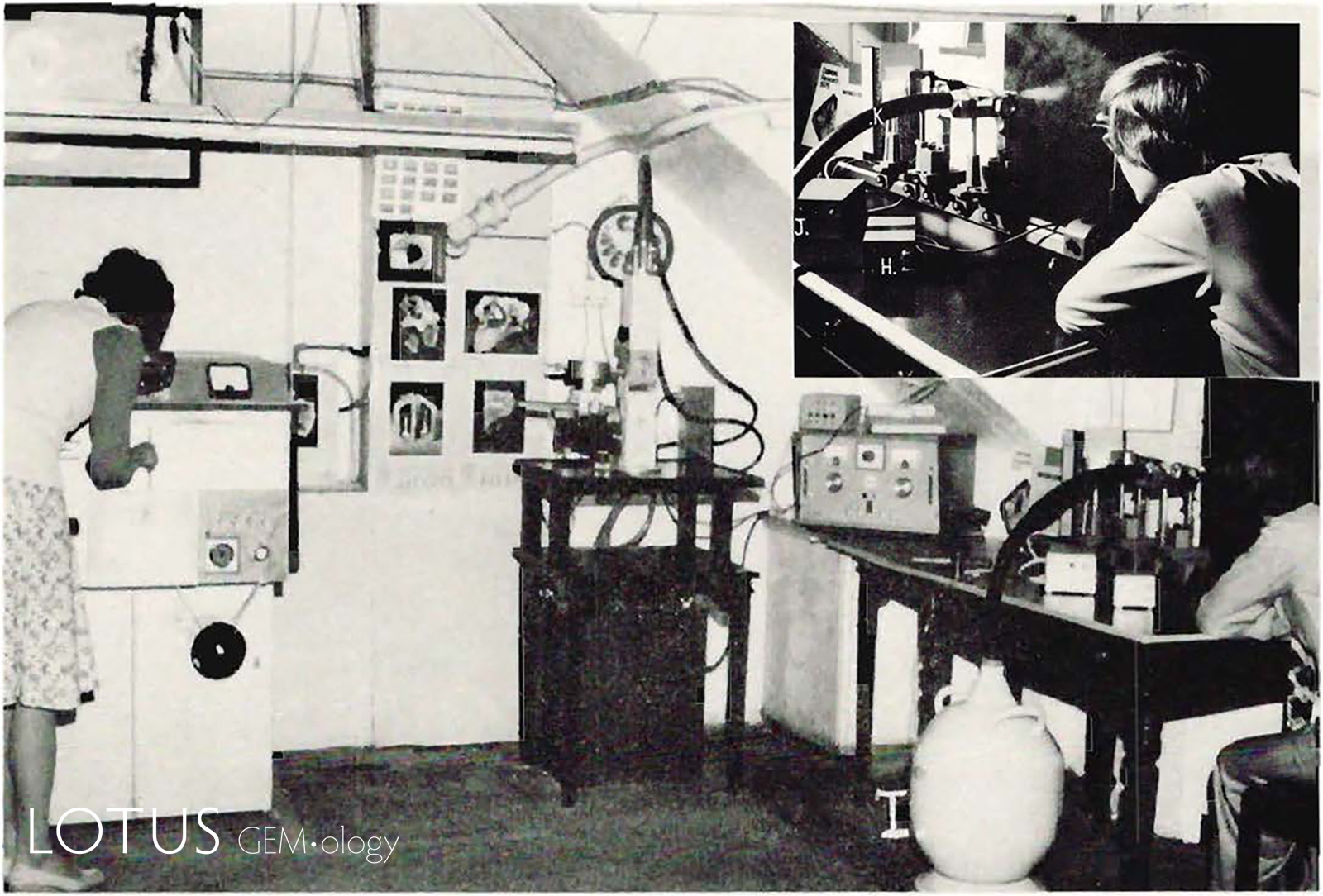 Figure 4. Section of the London Laboratory in the mid 1970's with a film based pearl X-ray unit to the left, a Debye-Scherrer X-ray Powder Diffraction (XRD) Camera system centrally positioned and low temperature spectroscopy set up for use with colored diamonds to the right of the image and inset top right.
The 1980’s saw FAX (facsimile) communications, which had been prevalent in the business world since the late 1960’s, finally adding to the ability of laboratories globally to communicate issues at speeds which were hitherto unknown. Now data was being passed between laboratories globally as soon as identification issues arose.
As the computerisation of instruments progress during the seventies and eighties so then gemological laboratories took advantage of the innovations by purchasing and employing, e.g., electron microscopes for imaging and chemistry (Cole & Monroe, 1967; Dunn, 1977; Hutchison et al., 1976; Sanders, 1985; Schwarz, 1981; Stockton & Manson, 1981; Wilson, 1972; Zussman, 1987) and eventually simpler EDXRF units to gain an idea of the chemistry of gem materials (Poirot,1992). The latter had become very useful in confirming visual observations made of titanium diffusion treatment in sapphires, particularly the so-call deep diffusion examples (Anonymous,1992; Brown, 1991; Crowningshield & Nassau, 1981; Fryer, 1991, 1993; R.W. Hughes,1988, 1992; Koivula & Kammerling, 1990–1992; Laughter, 1993; Lumetta, 1991; Petersen, 1992).
For a long time, infrared spectroscopy could not be applied to gemological studies as dispersive instruments required the partial or total destruction of the sample. At this time, the gem trade was facing issues for which potential solutions lay in the infrared. These included new types of hydrothermally grown synthetic emerald, plastic impregnated and synthetic opal, synthetic diamond, treated yellow diamonds which had been annealed to higher temperatures, amber identification issues and – critically for the colored gemstone world – the identification of heat-treated rubies and sapphires. Fortunately, just in time to meet the need and demand the Research Department of the Gemological Institute of America, then in Santa Monica, obtained a Nicolet SX Fourier Transform Infrared Spectrometer and published some excellent data (Fritsch et al. 1988; Fritsch and Shigley 1989; Shigley et al. 1987; Shigley and Fritsch 1989), the London Laboratory followed this with the purchase of a Nicolet 510 bench top model FTIR, here Scarratt pioneered the ‘misuse’ of the diffuse reflectance accessory’s alignment mirror to rapidly gain diamond typing data (Scarratt, 1989b) which while not providing quantifiable numbers did provide an understanding of a diamond’s nitrogen, hydrogen or boron content (Collins, 1978, 1982; Collins et al., 1989; Fritsch & Scarratt, 1989a–b, 1992–1993; Fritsch et al., 1991a–b; Lightowlers & Collins, 1976; Woods & Collins, 1983; Woods et al., 1990a–b; Woods et al., 1990a), Shortly after other laboratories added FTIR instruments to their gemological arsenal.
The late 1980’s saw the beginnings of a significant milestone in gemological education, research and gem testing laboratory activities within China, with Nelson, Jobbins and Scarratt coaching/teaching the first potential gemological instructors in Wuhan. Since these fledgling days, when reportedly there were only three qualified gemologists in China, today Chinese gemologists play a significant role in gemological education, research and testing activities (Nelson, 1990).
A particular issue in the 80’s and 90’s was with the prefusion of synthetic amethysts entering the market (Crowningshield, 1979; Lind et al., 1985; Samoylovich, 1982; Zecchini 1979a–b; Zecchini & Mérigoux, 1980; Zecchini & Smaali, 1999). This issue was thought at the time solvable with the observation of Brazil Law twinning in the natural material until the growth of synthetic material with the growth phenomena was suggested by Koivula & Fritsch (1989), Luckily systems were developed using FTIR that were able to identify, relatively inexpensively, the unscrupulous salting of parcels of natural amethyst with synthetics.
in 1981 Nassau wrote “with the ready availability of lasers, Raman scattering has now joined the growing list of non-destructive tests which can be applied to gemstones (Nassau, 1981) however it was not until 1994 that the Asian Institute of Gemological Sciences (AIGS) became the first laboratory to install Raman spectroscopy and deliver a presentation on how this could be used efficiently in the identification of fillers in emerald the same year (DuToit, 1995, 2009; Johnson et al., 1999; Nelson, 1993, 1995; Scarratt et al., 1996) the instrument installed in the AIGS laboratory was an early Renishaw instruments model. Other laboratories followed suit in quick succession as the importance of Raman spectroscopy and instrument related photoluminescence in gemology was clearly recognised.
By the mid 90’s gemological laboratories aided by instrument computerisation had evolved from establishments that employed what today are regarded as simple instruments (but were the backbone on which gemology developed), to a level of sophistication the that was impossible to visualise in 1925. But there was much more to come.
There was a great rapport between gemological laboratories in the early years where laboratory directors became close person friends, indeed during a conference in Lugano in 1952 Edward Gübelin proposed the creation of a “Committee of an International Gemmological Association” that would consist of one member per country; this member being the director of a gem testing laboratory, or a gemologist of the calibre who could attend that meeting. This was agreed to, and this meeting was later considered to be the inaugural meeting of the International Gemmological Conference (IGC). In 1987 the InterGemLab group of European Gemmological Laboratories was formed. Growing demands from the industry for treatment and origin determinations on a global level eventually led to a wider grouping under the heading of the Laboratory Manual Harmonisation Committee being formed in 2001 that initially brought together seven major international gemological laboratories based in the USA, Asia and Europe (Figure 5).
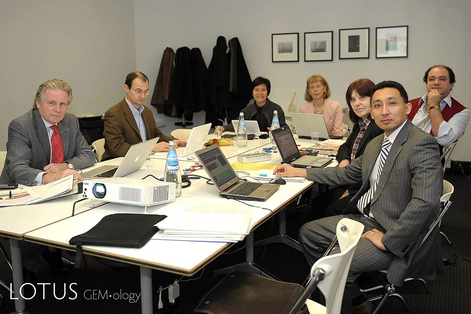 Figure 5. A 2010 meeting of LMHC members in Vicenza, Italy.
Following the introduction of computer assisted UV/visible/NIR, FTIR and Raman spectroscopy to the world of gemological laboratories, some innovative enthusiasts put their minds to the development of miniaturised versions that potentially could be easy to use for given applications. This made for greater efficiency and cost effectiveness in the approaching era of the commercially oriented gemological laboratory, rather than the academic approach that had previously prevailed. These new innovative instruments, while limited in their application when compared with their “research grade” counterparts, undoubtedly assisted in the efficiency and growth of the smaller gemological laboratories globally. Indeed in some establishments the refractometer became sidelined in favour using the miniaturised Raman spectrometer in the identification of gems (Hughes, E.B., 2015).
Edward Gübelin had pioneered distinguishing between differing gemstone origins producing and publishing in profusion photomicrographs that explained his approach in the use of inclusions as indicators as well as the identification of synthetic stones since the 1940’s (Gübelin, 1940a–b, d; Gübelin & Shipley, 1941; Gübelin, 1944a–c, 1973). These observations culminated in the publication of his books, Inclusions as a Means of Gemstone Identification (Gübelin, 1953) and twenty years later, the Internal World of Gemstones in 1973 (Gübelin, 1973). There then followed a collaboration between him and John Koivula, a later date gemstone inclusion enthusiast and avid practitioner of photomicrography that eventually resulted in The Photoatlas of Inclusions in Gemstones series (Gübelin & Koivula, 1986, 2005, 2008). In the early days of gemological photomicrography both Gübelin and Koivula used slow fine grained films to produce high resolution 35 mm transparencies with a camera system attached to a trinocular optical head. (Koivula 1986, 2003). As technology grew auto exposure systems were added.
Photomicrography excelled throughout the 80’s and 90’s as this medium laid bare the major and simple identification characteristics of new synthetic materials and treated gems for gemologists around the world (Hughes, R.W., 1987; Jobbins et al., 1976; Scarratt, 1976–1977, 1982, 1986a, 1987a–b, 1987d, 1988a–b,, 1989a; Scarratt & Harding, 1984; Scarratt et al., 1986).
 Figure 6. A Nikon SMZ25 digital photomicrography system with auto and fine adjustment focussing and optical fibre lighting, along with stacking software, in use at Danat in Bahrain in 2025. Photo Danat.
As digital alternatives to gemological photomicrography slowly became available “purists” dismissed this advance until significant improvements in resolution were found, plus the important availability of stacking software (Piper, 2010) that allowed for tremendous improvements in depth of field realisation. This new imaging became increasingly useful as a learning tool within laboratories particularly as that all the images could now be catalogued and stored for easy reference (Lotus, 2025).
Early in 2002, a significant turning point for gemological laboratories was reached, coincidentally with the use of e-mail as a primary communication method, as it became apparent that corundum treated by a new technique in Thailand had been filtering into the marketplace unannounced, particularly in Japan. The first announcement of this situation—an alert issued by the American Gem Trade Association (AGTA) on January 8, 2002—prompted substantial activity in gemological laboratories worldwide (Scarratt, 2002a–b). Quite rapidly it was demonstrated that this new process involved diffusion of the light element beryllium (Be) into a wide variety of corundum types to alter their color ( Emmett et al., 2003; McClure, 2005; Peretti & Günther, 2002; Peretti et al., 2003; DuToit, 2009;). However, the problem that now needed to be faced was that the EDXRF units used in gemological laboratories for analysing the trace element chemistry of gem materials could not detect beryllium, indeed the confirmation that beryllium was the diffused element was determined by Secondary-Ion Mass Spectrometry (SIMS), an instrument that was out of the purchasing power of even the best financed of gemological laboratories. Fortunately at this time LIBS spectroscopy (Ancillotti et al., 2004; D'Angelo et al., 2004; Hänni et al., 2004; Harmon et al., 2005; Krzemnicki et al., 2004; Rieger et al., 2002; Song et al., 2002; Tognoni et al., 2002) which if applied could detect the presence of Be, became available. However, initial issues surrounding laser damage to stones plus the discovery that Be could be present naturally in corundum led most major laboratories to embrace Laser Ablation Inductively Coupled Plasma Mass Spectrometry (LA-ICP-MS). This new tool not only could determine the presence of Be, it could also quantify the amount present in ppm. With this determination and correlation of the Be with other trace elements a conclusion could be made as to whether the Be was a product of a diffusion process or was naturally occurring.
Origin
A significant step for gemological laboratories related directly to the determination of origin, something that the trade used as an important marketing tool while serious laboratories regarded it as an academic exercise (the two viewpoints often resulting in a conflict) and complex treatments. Here in 2008 the Bangkok Laboratory of GIA enlisted Vincent Pardieu to set up and run the first properly managed Field Gemmology Department. Pardieu set about going to mine sites and collecting verifiable samples and cataloguing them in such a manner as to ensure the reliability of the samples and the data collected in perpetuity. Due to the extraordinary cost of such an enterprise the collecting and cataloguing of such a reliable data set had not previously been possible (Kautsky, 2016; Pardieu, 2009, 2020; Pardieu & Vertriest, 2016; Pardieu, Vertriest, Weeramonkhonlert, & Raynaud, 2017; Saeseaw, 2017; Staff 2015a–b; Vertriest, 2019). Such an enterprise with rigorously applied collection and cataloguing standards is a never-ending process as new gem deposits and well as alterations to old deposits continue unabated. The GIA recently announced the completion of their 100th field trip (GIA, 2025).
Origin and traceability determinations have recently taken on a different level of importance for the trade and laboratories as key jewellers and consumers appear to be increasingly focused on supply chain issues rather than values associated with origin (marketing), thus making such determinations important on a different platform. This has led to the development of ESG compliant trancing technology and AI machine learning using collected origin data.
Pearl Testing
By 2008 pearl testing using the film-based approaches developed in the early days of gemological laboratories were realised to be both laborious and inefficient, given the availability of modern technology. With the need for greater efficiencies and to train young gemologists to solve new issues that were arising between 2008 and 2020 gem testing laboratories involved in pearl testing began experimenting with real-time digital X-ray microradiography (RTX). This began when the Faxitron CS-100AC unit was installed in the GIA Laboratories in Bangkok and New York and after several iterations ended up with the more ergonomically friendly PXi GenX-90 which is high-resolution real-time X-ray unit that comes with a joystick powered x–y sample movement and flat panel detectors. The huge advantage with these units is that the high-resolution digital images could be stored and retrieved with a few clicks of the mouse rather than trying to sort through decades of film stored in brown envelopes (Al-Alawi, 2019; Karampelas, Al-Alawi, Al-Attawi, 2017; Scarratt & Karampelas, 2020).
Also developed in the 1960s, although not actively used in pearl laboratories until around 2010, X-ray μ-CT (computed X-ray microtomography) today allows well-funded pearl laboratories to explore, nondestructively, the internal structure of not just pearls but any object, with high spatial resolution. μ-CT units iteratively collect radiographic projections of a sample while it is rotating through 360°. Projections of the sample are recorded by a flat-panel detector with an integrated scintillator. The collected projections are used to construct three dimensional models of, in this case, the pearl. Then, two-dimensional (2D) slices can be cut through the 3D models in different directions allowing for the pearl’s internal structure to be examined slice by slice (Hainschwang, 2011; Karampelas et al., 2010; Krzemnicki et al., 2010; Scarratt & Karampelas, 2020).
Thus, the availability of RTX and μ-CT has allowed laboratories involved in pearl testing to make huge leaps in terms of the understanding and interpretation of growth structures, efficiencies, staff training and the reliability of results.
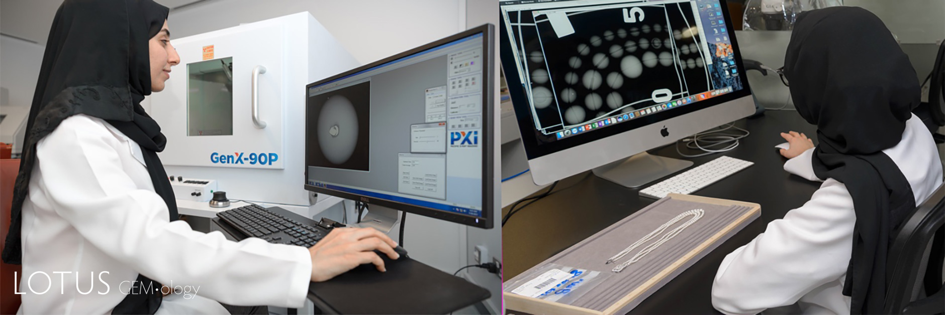 Figure 7: (left) an RTX unit for collecting micro X-ray imaging and (right) a work station for retrieving the micro X-ray imaging and making determinations on the nature of pearls examined in the Danat Laboratory.
A Century On
After 100 years of research and application it can be stated with a high degree of confidence that the words of Issac Newton “if I have seen further it is by standing on the shoulders of giants” are just as relevant today, for without the original research and applications developed for gemological laboratories in the 1920’s maybe we would not be at the advanced stage we are today. Where we will be in 100 years from now remains to be seen but for sure new and exciting innovations will emerge along with new and ever enthusiastic gemologists.
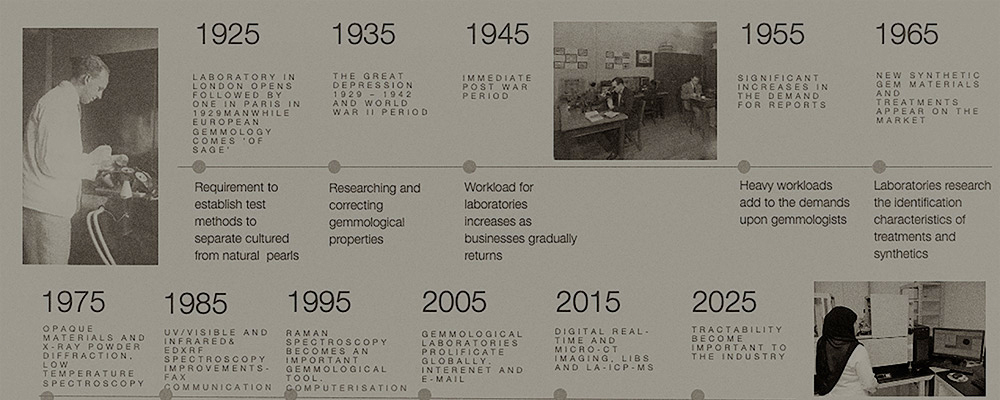
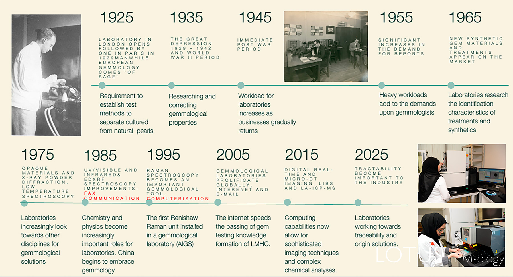 Figure 8. A graphic 100-year timeline outlining the development of gemological laboratories.
![]()
About the Author
Kenneth Scarratt is one of the world’s most notable gemmologists and an authority on pearls, having spent more than five decades researching and authenticating jewels, both the infamous and the secretly coveted. Kenneth has held management positions in prestigious international gemmological centres including the Gemological Institute of America, AGTA Gemological Testing Centre in New York, the Asian Institute of Gemological Sciences, Bangkok, DANAT and the Gemmological Association and Gem Testing Laboratory of Great Britain.
Footnotes
[1] Candling is a method used in embryology to study the growth and development of an embryo inside an egg. The method uses a bright light source behind the egg to show details through the shell and is so called because the original sources of light used were candles (wiki). In the case of pearls, the technique is used to reveal the layered growth bands of the shell beads used as the substrate for nacre growth in bead cultured pearls.
[2] Synthetic versions of gemstones are artificial products having essentially the same chemical composition, physical properties and structure as that of their naturally occurring counterparts.
[3] Benzyl benzoate is often used today for the examination of emerald in immersion as its RI is a close match to that of emerald, but as its viscosity is a little higher than, e.g., methylene iodide, that is a close match to with corundum, it is useful when examining ruby melee when laid out on a microscope slide.
[4] Located within the Square Mile of the City of London it is housed in two Grade-II listed buildings. It is a place packed with history – there has been a livestock market on the site for over 800 years – and yet it is thoroughly modern with its state-of-the-art facilities for the receiving, storing and despatching of meats.
Notes
First published in the Journal of Gems & Gemmology in May 2026.
References
- Al-Alawi., A., Ali., Z., Al Mahmood., F., Alderazi., H., Maklooq., F., Rajab., Z., Al Badel., F., Alatawi., A., Karampelas., S. (2019) Saltwater natural pearls from Pinctada radiata from the Kingdom of Bahrain. (Unpublished manuscript: DANAT).
- Alexander, A. E. and Sherwood, H. F. (1940) The radiography of cultured and natural pearls. The Gemmologist, Vol. 10, No. 113, pp. 45–48.
- Alexander, A.E (1941a) Natural and cultured pearl differentiation (cont…Radiography of natural and Cultured Pearls),' Gems & Gemology, Vol. 3, No. 12, pp. 185–188.
- Alexander, A.E. (1941b) Natural and cultured pearl differentiation (Part 1 and 2). Gems & Gemology, Vol. 3, No. 11/12, pp. 169–172 and 84–88.
- Ancillotti, L., Castellucci, E.M., and Becuccci, M. (2004) A combined Raman–LIBS spectrometer: Toward a mobile atomic and molecular analytical tool for in situ applications. SPIE Proceedings, No. 5850, pp. 182–189.
- Anderson, B.W. (1931) The use of X–rays in the study of pearls. British Journal of Radiology, Vol. 5, pp. 57–64.
- — (1935a) Igmerald. Gems & Gemology, Vol. 1, No. 10, pp. 284–285.
- — (1935b) "Synthetic diamonds". Gems & Gemology, Vol. 1, No. 8, pp. 213–216.
- — (1937a) Recent work on zircon: French Indo-China as the chief present-day source of zircon. The Gemmologist, Vol. 7, No. 74, pp. 611–612.
- — (1937b) Recent work on Zircon: The heat treatment of zircons. The Gemmologist, Vol. 7, No. 75, pp. 97–103.
- — (1944a) Gemstones and the spectroscope. Gems & Gemology, Vol. 4, No. 11, pp. 164–167.
- — (1944b) Gemstones and the spectroscope. Gems & Gemology, Vols 4, 5, Nos. 12, 1, pp. 180–181, 203–204.
- — (1950) Gemstones and the spectroscope – The absorption spectra of emerald and alexandrite. Gems & Gemology, Vol. 6, No. 9, pp. 263–266.
- — (1970) Some problems and a few solutions in the field of gem testing with the spectroscope. Gems & Gemology, Vol. 13, No. 8, pp. 238–244.
- — (1972) Spectroscope: An indicator of variation. Gems & Gemology, Vol. 14, No. 4, pp. 98–101.
- — (1973) 1925….and all that. Journal of Gemmology, Vol. 13, No. 7, pp. 249–262.
- — (1981) The Growning Pains of Gemmology. Journal of Gemmology, Vol. 17, No. 8, pp. 515–521.
- Anderson, B.W. and Payne, C.J. (1937a) Magnesium-zinc-spinels from Ceylon. Mineralogical Magazine, Vol. 25, No. 158, pp. 547–554.
- — (1937b) Recent work on zircon – III – Research in measurement of physical properties. The Gemmologist, Vol. 7, No. 77, pp. 297–301.
- — (1939a) An exceptional danburite. The Gemmologist, Vol. 8, No. 90, p. 105.
- — (1939b) The density of pearls and cultured pearls. The Gemmologist, Vol. 8, No. 94, pp. 166–169.
- — (1954) The spectroscope and its applications to gemmology. The Gemmologist, Vol. 23, No. 276, pp. 119–123.
- — (1955) The spectroscope and its applications to gemmology. The Gemmologist, Vol. 24, No. 285, pp. 68–71.
- — (1956) The spectroscope and its applications to gemmology: Part 35. The absoprtion spectrum of diamond. The Gemmologist, Vol. 25, No. 300, pp. 115–119.
- Anderson, B.W., Payne, C.J., and Pike, J. (1940) New refractometers employing diamond and other minerals. Mineralogical Magazine, Vol. 25, No. 170, pp. 579–583.
- Anderson, B.W., Payne, C.J. (1998), The Spectroscope and Gemmology, ed. Keith Mitchell R. (GemStone Press) p. 269.
- Anonymous (1992) Diffusion sapphires: Where do they go?. Jewellery News Asia, No. 89 (January), pp. 1,5.
- — (2024) Chelsea filter. Wikipedia.
- Bosshart, G. (1982) Distinction of natural and synthetic rubies by ultraviolet spectrophotometry. Journal of Gemmology, Vol. 18, No. 2, pp. 145–160.
- — (1989) The Dresden green. Journal of Gemmology, Vol. 21, No. 6, pp. 351–362.
- Brown, G. (1991) A new surface diffusion-treated sapphire. Australian Gemmologist, Vol. 17, No. 11, pp. 457–459.
- Burbage, E.J. and Anderson, B.W. (1942) An analysis of the movements of the shadow-edge on the refractometer in the case of biaxial gemstones. Mineralogical Magazine, Vol. 26, No. 178, pp. 246–253.
- Cole, S.H. and Monroe, E.A. (1967) Electron microscope studies of the structure of opal. Journal of Applied Physics, Vol. 38, No. 4, pp. 1872–1873.
- Collins, A.T. (1978) Investigating artificially coloured diamonds. Nature, Vol. 273, No. 5664, pp. 654–655.
- — (1982) Colour centres in diamond. Journal of Gemmology, Vol. 18, No. 1, pp. 37–75.
- Collins, A.T., Kamo, M., and Sato, Y. (1989) Optical centres related to nitrogen, vacancies and interstitials in polycrystalline diamond films grown by plasma–assisted chemical vapour deposition. Journal of Physics D: Applied Physics, Vol. 22, No. 9, pp. 1402–1405.
- Crowningshield, G.R. (1957) Spectroscopic recognition of yellow bombarded diamonds and bibliography of diamond treatment. Gems & Gemology, Vol. 9, No. 4, pp. 99–104, 117.
- — (1969) Orange-brown treated diamond. Gems & Gemology, Vol. 13, No. 3, pp. 89–90.
- — (1979) Synthetic amethyst. Gems & Gemology, Vol. 16, No. 5, pp. 151–152.
- Crowningshield, G.R. and Nassau, K. (1981) The heat and diffusion treatment of natural and synthetic sapphires. Journal of Gemmology, 17, No. 8, pp. 528–541.
- D'Angelo, C., et al. (2004) Spectroscopic analysis of signals from LIBS experiments. SPIE Proceedings, 5622, pp. 1037–1042.
- Dunn, P.J. (1977) The use of the electron microprobe in gemmology. Journal of Gemmology, Vol. 15, No. 5, pp. 248–258.
- DuToit, G., Thanachakaphad, J., Scarratt, K. (2009) Beryllium treated blue sapphires: Continuing market observations and update including the emergence of larger size stones. June 25th 2009.. News from Research.
- DuToit., G. (1995) Raman spectroscopy and its usefulness in identifying various internal features in gemstones.. International Gemmological Conference (Rayong, Thailand).
- Emmett, J., et al. (2003) Beryllium diffusion of ruby and sapphire. Gems & Gemology, Vol. 39, No. 2, pp. 84–135.
- Eppler, A. (1934), Edelsteine und Schmucksteine (Leipzig: Wilhelm Diebener) see pp. 319–343.
- Eppler, W.F. (1928) Die Edelsteinindustrie Siams [The gemstone industry of Siam] [in German]. Deutsche Goldschmiede-Zeitung.
- — (1933) Der Diamant und seine Bearbeitung. Deutsche Goldschmiede Zeitung, Vol. 22, No. 1, p. 62.
- — (1935) Mining emeralds in Colombia. The Gemmologist, Vol. 4, No. 43, pp. 201–207.
- — (1938) Die ideal-schleifformen durchsichtiger edelsteine. Zentralblatt fur Mineralogie, Geologie, und Palaontologie, Abteilung A, pp. 1–5.
- Fritsch, E. and Shigley, J.E. (1989) Contribution to the identification of treated colored diamonds: Diamonds with peculiar color-zoned pavilions. Gems & Gemology, Vol. 25, No. 2, pp. 95–101.
- Fritsch, E. and Scarratt, K.V.G. (1989a) Optical properties of one type of natural diamonds with high hydrogen content. 28th International Geological Congress, Workshop on Diamonds, pp. 21–22.
- Fritsch, E. and Scarratt, K. (1989b) Optical properties of some natural diamonds with high hydrogen content. in A. Feldman, Holly., S. (ed.), Diamond Optics II (S.P.I.E Proceedings Series, 1146; Bellingham, WA: Society for Photooptical Instrumentation Engineers), pp. 201–206.
- — (1992) Natural-color nonconductive gray-to-blue diamonds. Gems & Gemology, Vol. 28, No. 1, pp. 35–42.
- — (1993) Gemmological properties of type Ia diamonds with an unusually high hydrogen content. Journal of Gemmology, Vol. 23, No. 8, pp. 451–460.
- Fritsch, E., Scarratt, K., and Collins, A. (1991a) Optical Properties of Some Natural Diamonds with High Hydrogen Content. in R. Messier, Glass, J.T., Butler, J.E., Roy, R. (ed.), International Conference on New Diamond Science and Technology (Pittsburgh, PA.: Materials Research Society), pp. 671–676.
- — (1991b) Optical properties of diamonds with an unusually high hydrogen content. New Diamond Science and Technology, pp. 671–676.
- Fritsch, E., et al. (1988) Detection of treatment in two unusual green diamonds. Gems & Gemology, Vol. 24, No. 3, pp. 165–168.
- Fryer, C. (1991) Diffusion-treated sapphires in fine jewelry. Gems & Gemology, Vol. 27, No. 3, pp. 178–179.
- — (1993) Diffusion treatment obscured by mounting. Gems & Gemology, Vol. 29, No. 4, pp. 283–284.
- GIA (2025) GIA Field Gemology Team Completes One-Hundredth Expedition. Visit to Tanzania and Kenya marks 17 years of rigorous scientific study (https://hongkong.gia.edu/hk-gia-news-press/gia-field-gemology-team-completes-one-hundredth-expedition: GIA).
- Gübelin, E. and Shipley, R.M. (1941) The synthetic emerald. Gems & Gemology, Vol. 3, No. 10, pp. 146–150.
- Gübelin, E.J. and Koivula, J.I. (1986) Photoatlas of Inclusions in Gemstones. Zürich, Switzerland: ABC Edition, revised Jan. 1992; German edition, 1986 (Bildatlas der Einschlüsse Edelsteinen), 532 pp.
- Gübelin, E. J., Koivula, J. I. (2005), Photoatlas of Inclusions in Gemstones 2; Basel, Switzerland: Opinio).
- — (2008), Photoatlas of Inclusions in Gemstones 3; Basel, Switzerland: Opinio).
- Gübelin, E.J. (1939) Die Mineralien im Dolomit von Campolungo (Tessin). Schweizerische Mineralogische und Petrographische Mitteilungen, p. 19.
- — (1940a) Differentiation between Russian and Colombian emeralds. Gems & Gemology, Vol. 3, No. 6, pp. 89–92.
- — (1940b) Differences between Burma and Siam rubies. Gems & Gemology, Vol. 3, No. 5, pp. 69–72.
- — (1940c) Characteristics of Ceylon rubies. Gems & Gemology, Vol. 3, No. 8, pp. 121–124.
- — (1944a) Gemstone inclusions – Sapphire. Gems & Gemology, Vol. 4, No. 10, pp. 142–149.
- — (1944b) Gemstone inclusions – Emerald. Gems & Gemology, Vol. 4, No. 12, pp. 174–179.
- — (1944c) Gemstone inclusions – Ruby. Gems & Gemology, Vol. 4, No. 11, pp. 158–163.
- — (1953), Inclusions as a Means of Gemstone Identification (Los Angeles: GIA) 220 pp.
- — (1973), Internal World of Gemstones (reprinted 1983 edn.; Zürich: ABC Verlag) 234 pp.
- Hainschwang, T. (2011) Three-dimensional X-ray radiography – A new technique for gemstone and pearl testing. InColor, No. 16, Spring, pp. 30–34.
- Hänni, H.A., et al. (2004) Ein neues Instrument für die analytische Gemmologie: LIBS. Gemmologie – Zeitschrift der Deutschen Gemmologischen Gesellschaft, Vol. 53, No. 2/3, pp. 79–86.
- Harmon, R.S., et al. (2005) Laser-induced breakdown spectroscopy (LIBS) – An emerging field-portable sensor technology for real-time, in-situ geochemical and environmental analysis. 50th Annual Meeting, Electron Microscopy Society of America, Vol. 1, pp. 21–28.
- Hughes, E.B. (2015) Reviewing the GemmoRaman-532. InColor, No. 28, Spring, pp. 28–31.
- Hughes, R.W. (1988) Reappearance of surface-diffusion treated sapphires in Bangkok. ICA Lab Alert, (12).
- — (1992) Devil’s Advocate: Vampire blues: deep-diffusion treated sapphires. JewelSiam, Vol. 3, No. 3, pp. 83–86.
- Hughes, R.W. . (1987) Glass infilling of cracks in ruby.. ICA Lab Alert, (4), 1.
- Hutchison, J.S., et al. (1976) Structural irregularities in nephrite jade: An electron microscope study. Materials Research Bulletin, Vol. 11, No. 12, pp. 1557–1562.
- Jobbins, E.A., Statham, P.M., and Scarratt, K. (1976) Internal structures and identification of Gilson synthetic opals. Journal of Gemmology, Vol. 15, No. 2, pp. 66–75.
- Johnson, M.L., Elen, S., and Muhlmeister, S. (1999) On the identification of various emerald filling substances. Gems & Gemology, Vol. 35, No. 2, pp. 82–107.
- Karampelas, S., et al. (2010) X-ray computed microtomography applied to pearls: Methodology, advantages and limitations. Gems & Gemology, Vol. 46, No. 2, pp. 122–127.
- Karampelas, S., Al-Alawi, A.T., Al-Attawi, A. (2017) Real-time Microradiography of Pearls: A comparison Between Detectors. Gems & Gemology, Vol. 53, No. 4, pp. 452–456.
- Kautsky, J., (2016) Pardieu Brings Ruby Rush to Life with Stories, Updates from Madagascar'. GIA.
- Kiefert, L. and Schmetzer, K. (1991a) The microscopic determination of structural properties for the characterization of optical uniaxial natural and synthetic gemstones – Part 3: Examples for the applicability of structural features for the distinction of natural and synthetic sapphire, ruby, amethyst and citrine. Journal of Gemmology, Vol. 22, No. 8, pp. 471–482.
- — (1991b) The microscopic determination of structural properties for the characterization of optical uniaxial natural and synthetic gemstones – Part 1: General considerations and description of the methods. Journal of Gemmology, Vol. 22, No. 6, pp. 344–354.
- — (1991c) The microscopic determination of structural properties for the characterization of optical uniaxial natural and synthetic gemstones – Part 2: Examples for the applicability of structural features for the distinction of natural emerald from flux-grown and hydrothermally-grown synthetic. Journal of Gemmology, Vol. 22, No. 7, pp. 427–438.
- Koivula, J.I. (1982) Inclusions in a better light. Edited by Eash, D.M., In International Gemological Symposium Proceedings, Santa Monica, CA: Gemological Institute of America, pp. 471–476.
- Koivula, J.I. (1986) Photomicrography – A "how-to" for today's jeweler-gemologist. The Scope, Vol. 2, No. 1, pp. 1–3.
- — (2003) Photomicrography for gemologists. Gems & Gemology, Vol. 39, No. 1, pp. 4–23.
- Koivula, J.I. and Fryer, C.W. (1984) Lepidolite with simulated matrix. Gems & Gemology, Vol. 20, No. 1, pp. 42–44.
- Koivula, J.I. and Fritsch, E. (1989) The growth of Brazil-twinned synthetic quartz and the potential for synthetic amethyst twinned on the Brazil law. Gems & Gemology, Vol. 25, No. 3, pp. 159–164.
- Koivula, J.I. and Kammerling, R.C. (1990) Diffusion-treated sapphire update. Gems & Gemology, Vol. 26, No. 4, p. 307.
- — (1991) More experimentation with blue diffusion-treated sapphires. Gems & Gemology, Vol. 27, No. 3, pp. 187–188.
- Koivula, J.I. and Kammerling, R. (1992) Diffusion treatment: How to do it and how to detect it. Jewellry News Asia, , No. 89, pp. 56–58.
- Krzemnicki, M., et al. (2010) X-ray computed microtomography: Distinguishing natural pearls from beaded and non-beaded cultured pearls. Gems & Gemology, Vol. 46, No. 2, pp. 128–134.
- Krzemnicki, M.S., Hänni, H.A., and Walters, R.A. (2004) A new method for detecting Be diffusion-treated sapphires: Laser-Induced Breakdown Spectroscopy (LIBS). Gems & Gemology, Vol. 40, No. 4, pp. 314–322.
- Larsen, E.S. (1921) The Microscopic Determination of the Nonopaque materials. in Department of the Interior (ed.), (Washington: Government Printic office).
- Laughter, T. (1993) Diffusion illusions. JewelSiam, pp. 62–65.
- Liddicoat, R.T. (1984) Basil W. Anderson 1901–1984. Gems & Gemology, Vol. 20, No. 1, p 1.
- Liddicoat, R.T. and Ball, S.H. (1941) The mining of gems and ornamental stones by American Indians. Gems & Gemology, Vol. 3, No. 12, pp. 178–181.
- Lightowlers, E.C. and Collins, A.T. (1976) Boron measurements in natural semiconducting diamond. Diamond Research, pp. 14–21.
- Lind, T., Schmetzer, K., and Bank, H. (1983) The identification of turquoise by infrared spectroscopy and X-ray powder diffraction. Gems & Gemology, Vol. 19, No. 3, pp. 164–168.
- — (1985) Methods for the distinction of natural and synthetic amethysts. Australian Gemmologist, Vol. 15, No. 12, pp. 462–470.
- Lotus (2025), Hyperion, lotus gemology inclusion search engine.
- Lumetta, P. (1991) Diffusion confusion. Gemological Digest, Vol. 3, No. 2, pp. 32–33.
- McClure, S.F. (2005) Le traitement des corindons au béryllium: Une mise au point. Revue de Gemmologie a.f.g., , No. 153, pp. 4–7.
- Mitchell, R.K. (1980) Anderson on heavy liquids . Journal of Gemmology, Vol. 17, No. 4, pp. 230–235.
- Nassau, K. (1981) Raman spectroscopy as a gemstone test. Journal of Gemmology, Vol. 17, No. 5, pp. 306–320.
- Nelson, J.B. (1960) The Debye-Scherrer powder camera. X-ray Diffraction of Polycrystalline Materials, pp. 78–121.
- — (1985) Nelson gemstone immersion microscope. Nelson Gemmological Instruments, pp. 1–14.
- — (1990) Gemmological teaching in Hong Kong and China. Journal of Gemmology, Vol. 22, No. 4, pp. 224–232.
- — (1991) Gemmological teaching in Catalonia. Journal of Gemmology, Vol. 22, No. 6, pp. 337–343.
- — (1993) La Microsonde Raman en Gemmologie. Mineralogical Magazine, Vol. 57, No. 389, pp. 763–766.
- — (1995) The identification of subsurface inclusions with laser Raman spectroscopy. 25th International Gemmological Conference – Abstracts, pp. 1–23.
- Nelson., J.B. (1995) Identification of subsurface inclusions with Raman spectroscopy. International Gemmological Conference (Rayong Thailand).
- Pardieu, V. (2009) Melos and their Pearls in Vietnam (May‐June 2009). Concise Field Report (GIA.edu: GIA Laboratory Bangkok).
- — (2020) Field Gemology, the evolution of data collection. InColor, , No. 46, pp. 36–42.
- Pardieu, V., Vertriest, W. (2016) Gem News International – Update on colored gemstone mining in Tanzania.. Gems & Gemology, Vol. 52, No. 3, pp. 318–321.
- Pardieu, V., Vertriest, W., Weeramonkhonlert, V., Raynaud, V., Atikarnsakul, U. and Perkins, R. (2017) Sapphires from the gem rush Bemainty area, Ambatondrazaka (Madagascar). GIA News from Research, 26 February, 45 pp.
- Peretti, A. and Günther, D. (2002) Color enhancement of natural fancy sapphires with a new heat-treatment technique.. Contributions to Gemology, No. 1, pp. 1–48.
- Peretti, A., Günther, D., and Graber, A. (2003) The beryllium treatment of fancy sapphires with a new heat-treatment technique (Part B).. Contributions to Gemology, No. 2, pp. 21–33.
- Petersen, G.E. (1992) Surface diffusion sapphires. Jewellery Time, No. 5, pp. 18–19.
- Piper, Jörg (2010) Software-based stacking techniques to enhance depth of field and dynamic range in digital photomicrography. in Tim D. Hewitson and Ian A. Darby (eds.), Histology Protocols (Totowa, NJ: Humana Press), pp. 193–210.
- Poirot, J.P. (1992) Spectrométrie et fluorescence X, des aides pour la détermination de types de gisement de saphirs. Revue de Gemmologie a.f.g.,No. 110), pp. 7–9.
- Poirot, J.P. and Gonthier, E. (1998) Le controle des perles a partir de 1929 au Laboratoire Gemmologique Francais (du laboratoire syndical au laboratoire C.C.I.P.). Revue de Gemmologie a.f.g., No. 133), pp. 26–27.
- Rieger, G.W., et al. (2002) Laser-induced breakdown spectroscopy for microanalysis using submillijoule UV laser pulses. Applied Spectroscopy, Vol. 56, No. 6, pp. 689–698.
- Saeseaw, S., Sangsawong, S., Vertriest, W., Atikarnsakul, U., Raynaud-Flattot, V., Khowpong, C., Weeramonkhonlert, V. (2017) A Study of Sapphires from Chantaburi, Thailand and it’s Gemological Characteristics. GIA News from Research.
- Samoylovich, M.I. (1982) Optical spectrum characteristics of synthetic amethyst crystal. International Geology Review, Vol. 24, No. 1, 41–43.
- Sanders, J.V. (1985) Structure of opals. Journal de Physique, Vol. 46, No. 3, pp. C3–1–C3–8.
- Scarratt, K (2002) Further Characterization of Sapphires Recently Treated in Bangkok. <http://www.agta.org/consumer/gtclab/treatedsapps01.htm>, accessed Friday, April 19.
- — (2002) AGTA and GIA Researchers Meet with Thai Treaters. <http://www.agta.org/consumer/gtclab/thaitreatermtg.htm>, accessed Monday, March 18, 2002.
- Scarratt, K. (1976) Notes on Gilson synthetic white opal (September 1975). Journal of Gemmology, Vol. 15, No. 2, pp. 62–75.
- — (1977) A study of recent Chatham synthetic ruby and synthetic blue sapphire crystals with a view to the identification of possible faceted material. Journal of Gemmology, Vol. 15, No. 7, pp. 347–353.
- — (1979) Investigating the visible spectra of coloured diamonds. Journal of Gemmology, Vol. , No. 7, pp. 433–447.
- — (1982) How to recognize the new Seiko synthetics. Retail Jeweller.
- — (1984) Notes from the laboratory. Journal of Gemmology, Vol. 19, No. 2, pp. 98–124.
- — (1986a) Synthetic opal. Journal of Gemmology, Vol. 20, No. 2, pp. 93–95.
- — (1986b) Notes from the Laboratory: Cape series at low temperatures. Journal of Gemmology, Vol. 20, No. 4. pp. ??
- — (1987a) Notes from the Laboratory: Biron synthetic emerald. Journal of Gemmology, Vol. 20, No. 5. pp. ??
- — (1987b) Russian hydrothermal synthetic emeralds. Journal of Gemmology, Vol. 20, No. 7/8, pp. 412–420.
- — (1987c) Notes from the Laboratory: Saussurite. Journal of Gemmology, Vol. 20, No. 6, pp. ??
- — (1987d) Glass infilling of cavities in ruby and sapphire. Journal of Gemmology, Vol. 20, No. 7/8, p. 421.
- — (1988a) Notes from the Laboratory: Lennix synthetic emerald. Journal of Gemmology, Vol. 21, No. 3, pp. ??
- — (1988b) Notes from the Laboratory: Glass filled feathers and cavities in ruby. Journal of Gemmology, Vol. 21, No. 3, pp. ??
- — (1989a) Notes from the Laboratory: DeBeers and Sumitomo synthetic diamonds. Journal of Gemmology, Vol. 21, No. 6, pp. ??
- — (1989b) Notes from the Laboratory – 14. Journal of Gemmology, Vol. 21, No. 6, pp. 339–346.
- Scarratt, K. and Harding, R.R. (1984) Glass infilling of cavities in natural ruby. Journal of Gemmology, Vol. 19, No. 4, pp. 293–297.
- Scarratt, K., Harding, R.R., and Din, V. K. (1986) Glass fillings in sapphire. Journal of Gemmology, Vol. 20, No. 4, pp. 203–207.
- Scarratt, K., Dunaigre, C.M., and DuToit, G. (1996) Chatham synthetic diamonds. Journal of the Gemmological Association of Hong Kong, Vol. 19, pp. 6–12.
- Scarratt K., (2002) Orange-pink sapphire alert'. American Gem Trade Association <http://www.agta.org/consumer/gtclab/orangesap-phirealert.htm.>
- Scarratt, K., Karampelas, S. (2020) Pearl testing and the historic use of X-rays. InColor, (Spring/Summer), pp. 82–86.
- Schwarz, D. (1981) Edelsteinlagerstättenforschung und Rasterelektronenmikroskop. Uhren Juwelen Schmuck, Vol. 20.
- Shigley, J.E. and Fritsch, E. (1989) Comparison of natural and synthetic diamonds. 28th International Geological Congress, Workshop on Diamonds, pp. 96–99.
- Shigley, J.E., et al. (1987) The gemological properties of the De Beers gem-quality synthetic diamonds. Gems & Gemology, Vol. 23, No. 4, pp. 187–206.
- Shipley, R.M. and Liddicoat, R.T. (1941) A solution to diamond color grading problems. Gems & Gemology, Vol. 3, No. 11, pp. 162–167.
- Smith, G.F.H. (1913), Gem-Stones and their Distinctive Characters (2nd edition (1st ed. 1912) edn.; London: Methuen & Co., 312 pp.
- Song, K., Lee, Y., and Sneddon, J. (2002) Recent developments in instrumentation for laser induced breakdown spectroscopy. Applied Spectroscopy Reviews, Vol. 37, No. 1, pp. 89–117.
- Staff, (2015a) GIA Field Gemologist Documents Madagascar Ruby Rush'.
- —, (2015b) GIA Field Gemology Team Explores Sapphire Mines at Ilakaka, Madagascar'. <https://www.gia.edu/gia-news-research/sapphire-mines-ilakaka-madagascar-field-expedition>
- Stockton, C.M. and Manson, D.V. (1981) Scanning electron microscopy in gemology. Gems & Gemology, Vol. 17, No. 2, pp. 72–79.
- Tognoni, E., et al. (2002) Quantitative micro-analysis by laser-induced breakdown spectroscopy: A review of the experimental approaches. Spectrochimica Acta B, Vol. 57, No. 7, pp. 1115–1130.
- Vertriest, W., Palke, A.C., Renfro, N.D. (2019) Field Gemology: Building a Research Collection and Understanding the Developments of Gem Deposits. Gems & Gemology, Vol. 55, No. 4, pp. 490–511.
- Webster, R (1955) X-rays and Their Use in Gemmology: Part V: Laue Patterns. The Gemmologist, Vol. , No. 289, pp. 148–151.
- Webster, R. (1957) The Detection of Cultured Pearls: Part 1: How it started. The Gemmologist, Vol. 26, No. 315, pp. 178–184.
- Wilson, W.E. (1972) The state of the art the electron microscopes and electron microprobe. Mineralogical Record, Vol. 3, No. 4, pp. 151–164.
- Woods, G.S. and Collins, A.T. (1983) Infrared absorption spectra of hydrogen complexes in type I diamonds. Journal of Physics and Chemistry of Solids, Vol. 44, No. 5, pp. 471–475.
- — (1986) New developments in spectroscopic methods for detecting artifically coloured diamonds. Journal of Gemmology, Vol. 20, No. 2, pp. 75–82.
- Woods, G.S., van Wyk, J.A., and Collins, A.T. (1990a) The nitrogen content of type Ib synthetic diamond. Philosophical Magazine B, Vol. 62, No. 6, pp. 589–595.
- Woods, G.S., et al. (1990b) The nitrogen content of type Ia natural diamonds. Journal of Physics and Chemistry of Solids, Vol. 51, No. 10, pp. 1191–1197.
- Zecchini, P. (1979a) Une méthode pour reconnaître les quartz synthétiques étude de l'absorption infrarouge de quartz d'origine naturelle ou de synthèse. Revue de Gemmologie a.f.g., No. 60, pp. 14–18.
- — (1979b) Étude de l'absorption infrarouge de quartz d'origine naturelle ou de synthése. Revue de Gemmologie a.f.g., No. 60, pp. 14–18.
- Zecchini, P. and Mérigoux, H. (1980) Étude de l'absorption infrarouge des quartz hyalin et colorés, naturels ou de synthese: application a la gemmologie. Comptes Rendus Hebdomadardies des Seances de l'Academie des Sciences D, pp. 290, 291–294.
- Zecchini, P. and Smaali, M. (1999) Identification de l'origine naturelle ou artificielle des quartz. Revue de Gemmologie a.f.g., Nos. 138–139, pp. 74–83.
- Zhou, C. (2021) A Brief History of Pearl Testing through the pages of Gems & Gemology. Journal of the Gemmological Association of Hong Kong, Vol. 42, pp. 88–95.
- Zussman, J. (1987) Minerals and the electron microscope. Mineralogical Magazine, Vol. 51, pp. 129–138.
![]()