Sapphire Inclusions
Explore sapphire inclusions in Hyperion, the Lotus Gemology inclusion database. Hyperion contains photomicrographs of inclusions and internal features in natural sapphire, documented with information on geographic origin, treatment, lighting conditions, field of view, photographer and published references.
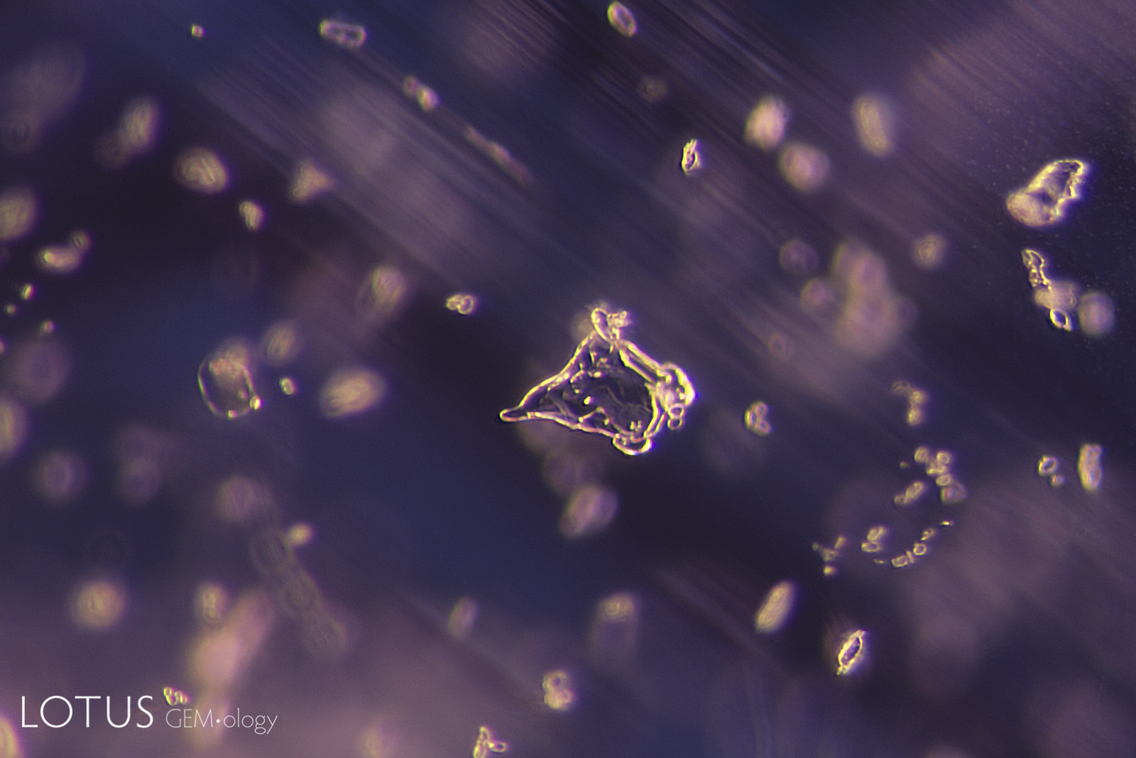
A common guest mineral in sapphire is apatite. These can take on a variety of forms, from rounded balls, such as that shown here, to hexagonal prisms and dog’s tooth shapes. This example was identified as fluorapatite by micro Raman.
Natural Sapphire •
Myanmar (Burma); Mogok •
Enhancements:
None Detected (None) •
Lighting Conditions:
Dark Field
Photographer:
Richard W. Hughes •
Image Number:
A-003-6364-3
• Field of View = 2mm
Hughes, R.W., Manorotkul, W. and Hughes, E.B. (2017) Ruby & Sapphire: A Gemologist's Guide. Bangkok, Lotus Publishing, 816 pp.; RWHL*.
Gübelin, E.J. and Koivula, J.I. (2008) Photoatlas of Inclusions in Gemstones, Volume 3. Basel, Switzerland, Opinio Publishers, 672 pp.; RWHL*.
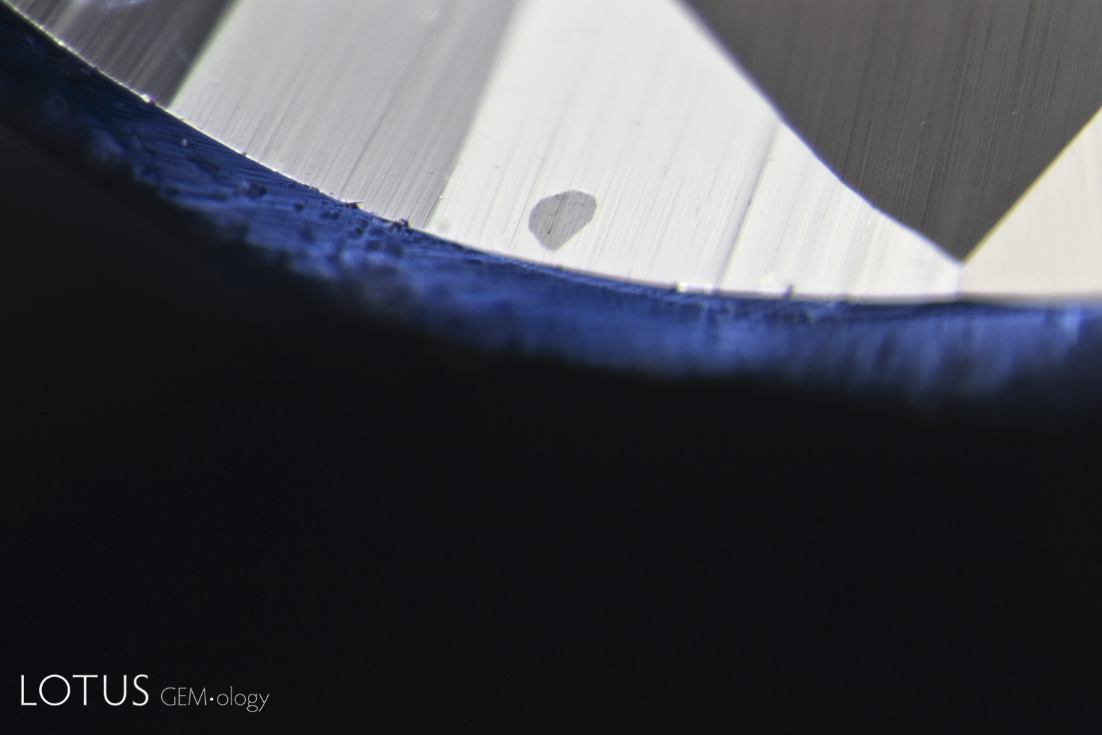
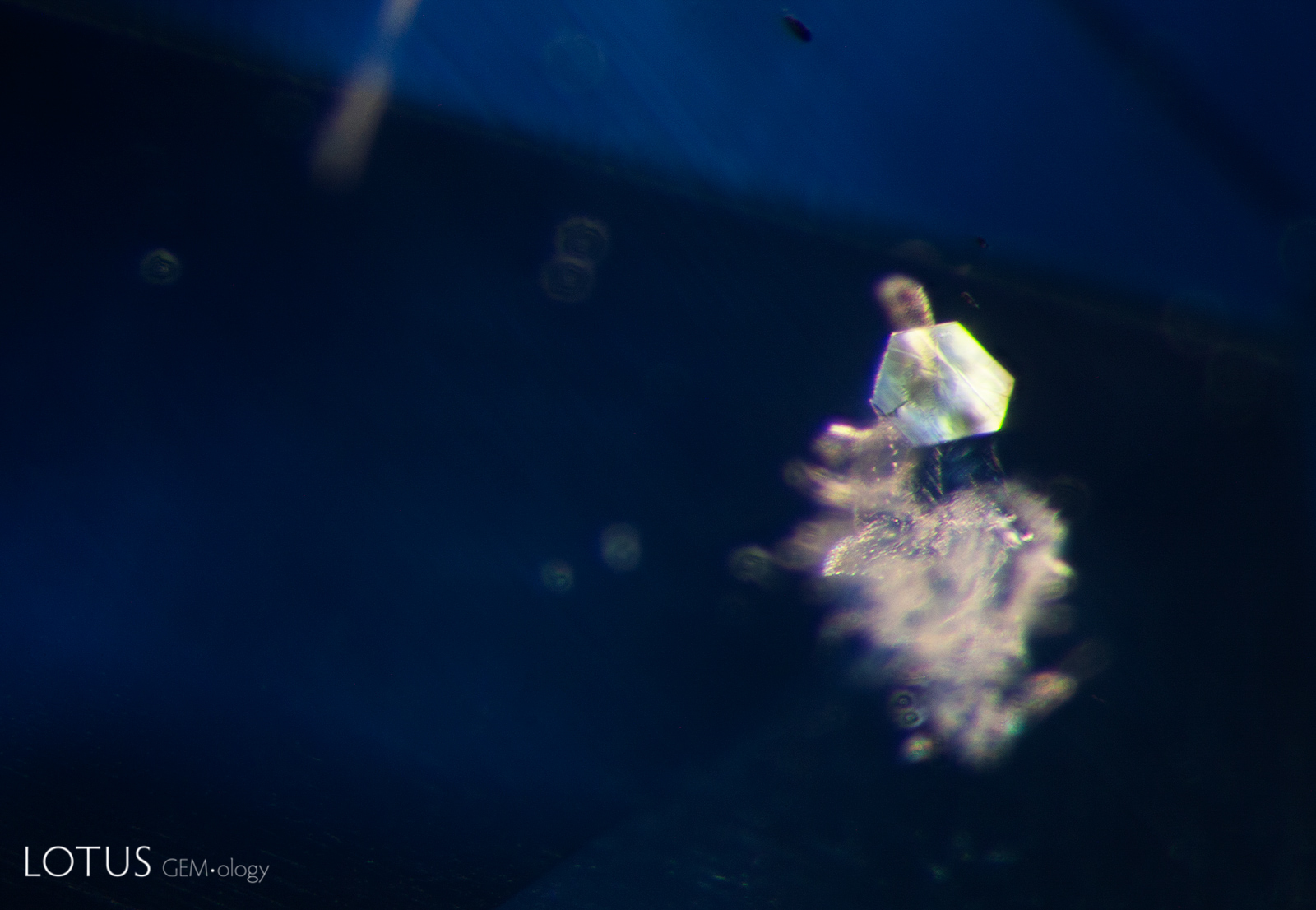
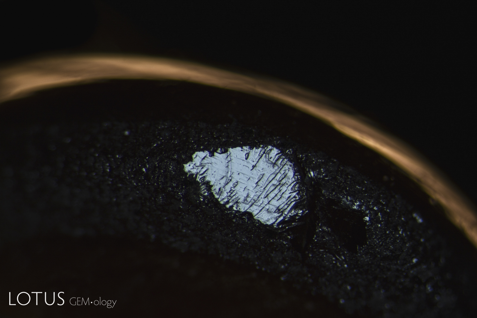
Two small sapphires were submitted to Lotus Gemology for testing. Both were said to come from Kashmir (Sumjam, India). One of the stones had a small transparent crystal grain cut through on the surface, as shown in the center of this photo. Using micro Raman, we identified the inclusion as anorthite feldspar. Feldspars are common in Kashmir sapphire. On the other hand, apatite has not been reported in Kashmir sapphire, but it is common in Sri Lankan and Madagascar sapphires.
Natural Sapphire •
India (Kashmir) •
Enhancements:
None Detected (None) •
Lighting Conditions:
Fiber Optic: Diffuse Overhead
Photographer:
Richard W. Hughes •
Image Number:
A-003-6342-1
• Field of View = 1.2mm
Hughes, R.W., Manorotkul, W. and Hughes, E.B. (2017) Ruby & Sapphire: A Gemologist's Guide. Bangkok, Lotus Publishing, 816 pp.; RWHL*.
Hänni, H.A. (1990) A contribution to the distinguishing characteristics of sapphire from Kashmir. Journal of Gemmology, Vol. 22, No. 2, pp. 67–75; RWHL*.
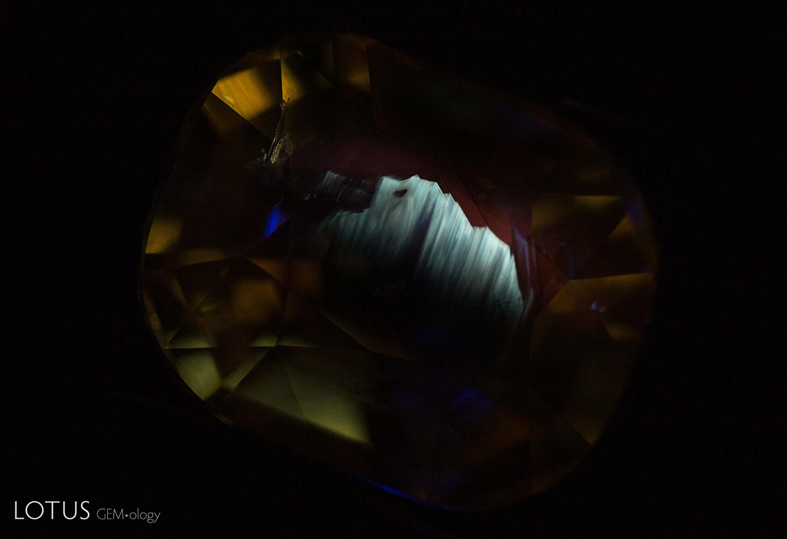
This yellow sapphire displays a striking angular, superficial chalky patch when viewed in shortwave UV illumination. This is the first time we have seen this reaction in a yellow sapphire that shows no signs of heat treatment, with all inclusions (intact negative crystals) and spectral information (strong 3160 in the infrared) pointing to an untreated gem. We have seen this reaction occasionally in untreated blue sapphires from Madagascar.
Natural Sapphire •
Sri Lanka (Ceylon) •
Enhancements:
None Detected (None) •
Lighting Conditions:
Ultraviolet: Shortwave
Photographer:
E. Billie Hughes •
Image Number:
A-003-6345-1
Hughes, R.W. and Emmett, J.L. (2005) Heat Seeker: UV fluorescence as a gemological tool. The Guide, Vol. 24, No. 5, Part 1, Sept.–Oct., pp. 1, 4–7; RWHL*.
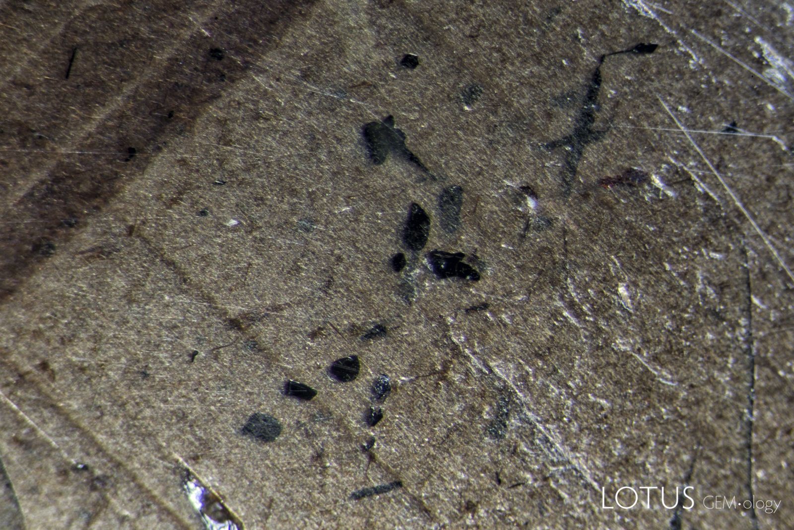
This hexagonal crystal with a metallic luster stood out in its sapphire host. Micro Raman analysis confirmed its identity as graphite.
Natural Sapphire •
Madagascar •
Enhancements:
None Detected (None) •
Lighting Conditions:
Dark Field + Diffuse Fiber Optic
Photographer:
E. Billie Hughes •
Image Number:
A-003-6344-1
• Field of View = 2.2mm
Hughes, R.W., Manorotkul, W. and Hughes, E.B. (2017) Ruby & Sapphire: A Gemologist's Guide. Bangkok, Lotus Publishing, 816 pp.; RWHL*.
Gübelin, E.J. and Koivula, J.I. (2008) Photoatlas of Inclusions in Gemstones, Volume 3. Basel, Switzerland, Opinio Publishers, 672 pp.; RWHL*.
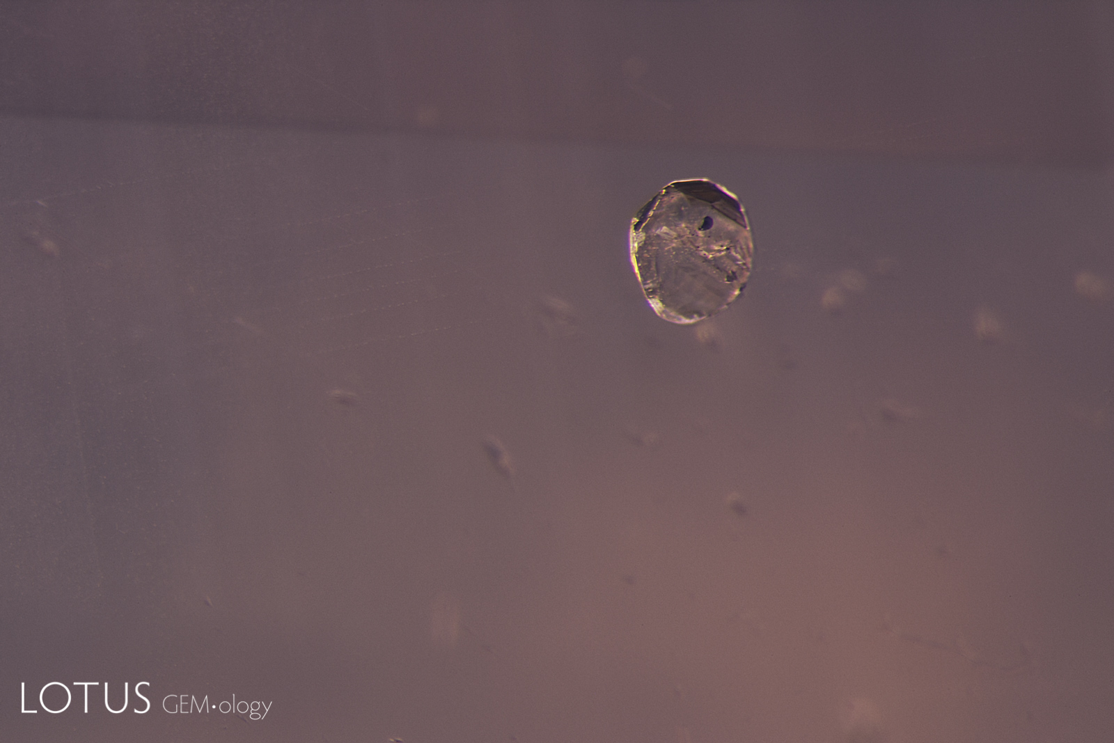
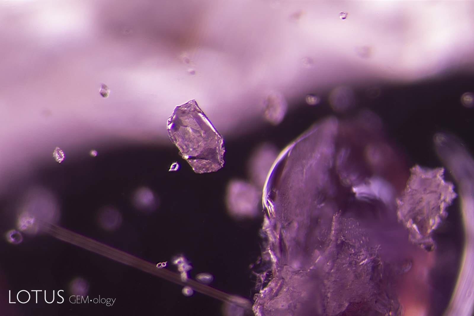
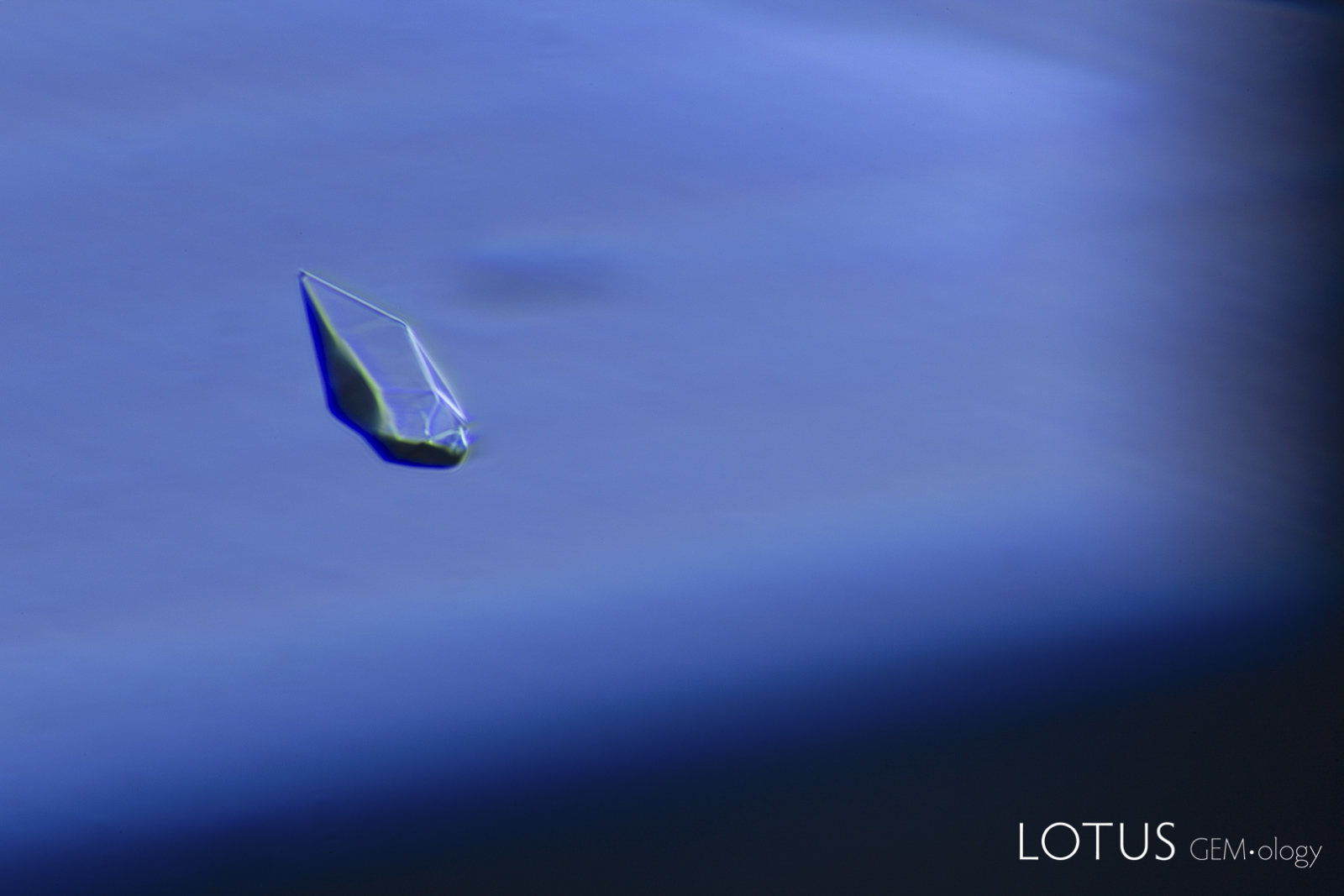
This untreated pink sapphire from the Ilakaka area of Madagascar was part of a large parcel purchased from a miner by the author in 2005. It contains zoned clouds of fine rutile dust, rounded zircon crystals, and in the center, an irregular rounded crystal that was identified by micro Raman as anorthite, one of the end members of the plagioclase feldspar series.
Natural Sapphire •
Madagascar •
Enhancements:
None Detected (None) •
Lighting Conditions:
Dark Field
Photographer:
Richard W. Hughes •
Image Number:
A-003-6353-1
• Field of View = 2mm
Hughes, R.W., Manorotkul, W. and Hughes, E.B. (2017) Ruby & Sapphire: A Gemologist's Guide. Bangkok, Lotus Publishing, 816 pp.; RWHL*.
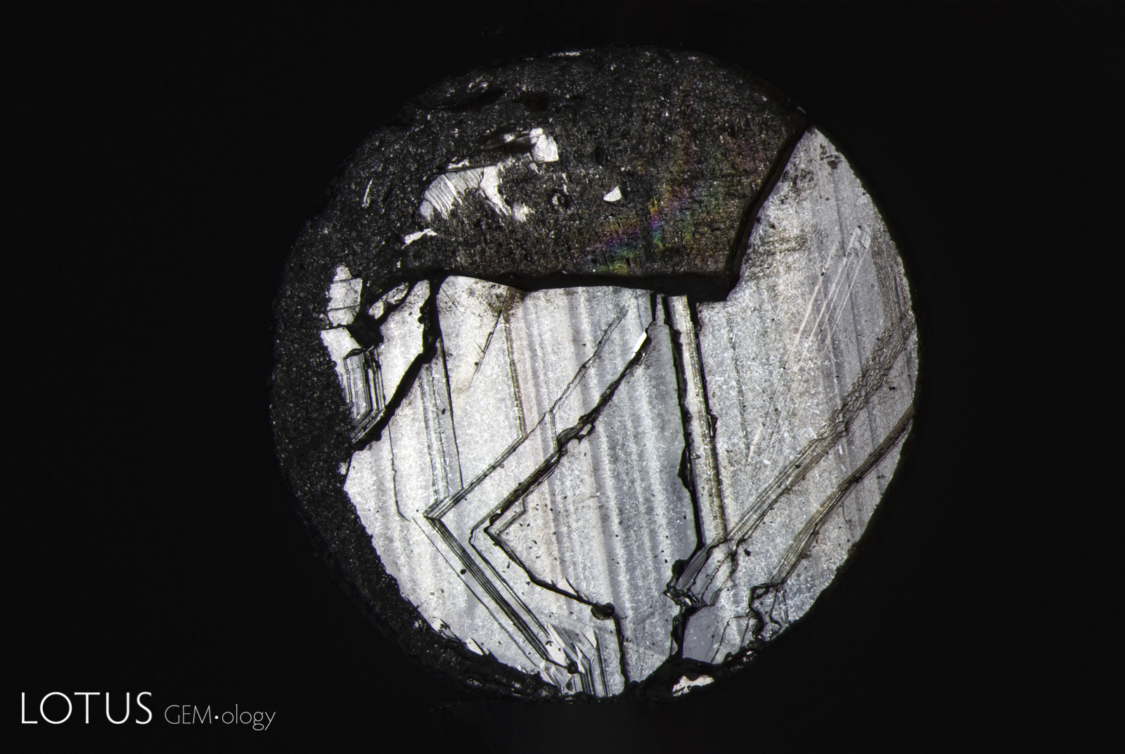
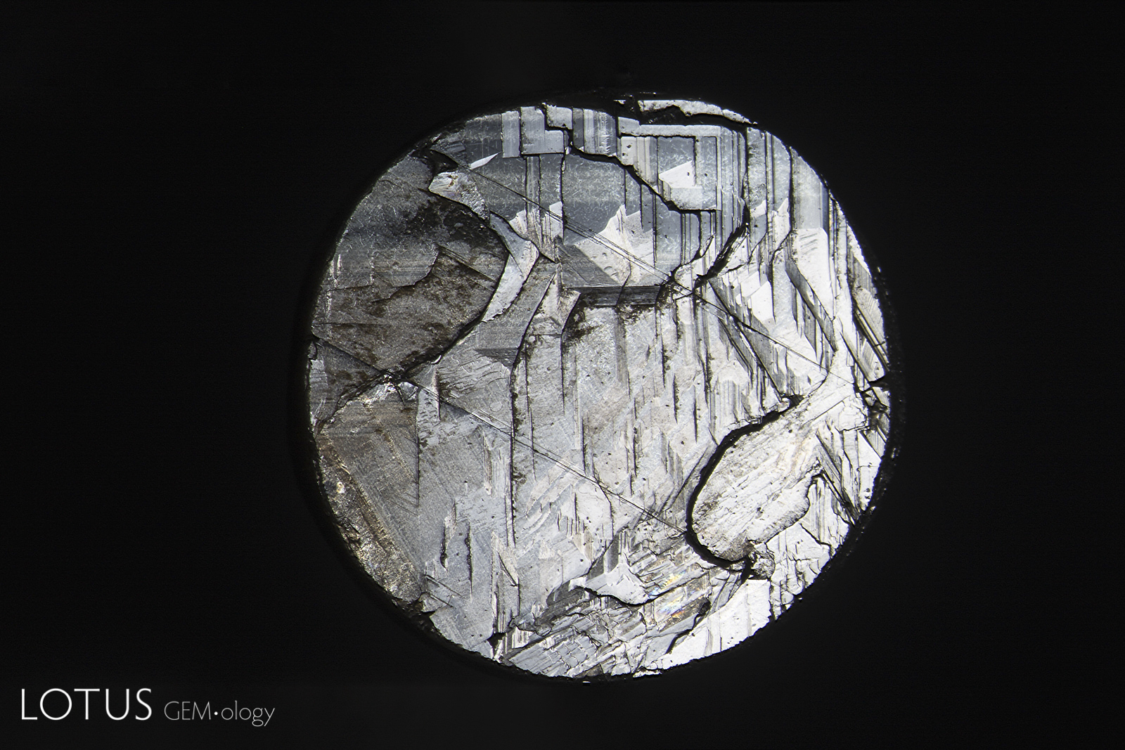
Basal parting seen on the back of a black star sapphire from Chanthaburi, Thailand. Note the step-like appearance. The two diagonal lines are rhombohedral parting planes. Basal parting in these stones is due to exsolution of hematite/ilmenite silk, which produces both the star effect and a lack adhesion.
Natural Black Star Sapphire •
Thailand (Siam); Chanthaburi •
Enhancements:
None Detected (None) •
Lighting Conditions:
Fiber Optic: Diffuse Overhead
Photographer:
Richard W. Hughes •
Image Number:
R-003-6291-1
• Field of View = 11mm
Hughes, R.W., Manorotkul, W. and Hughes, E.B. (2017) Ruby & Sapphire: A Gemologist's Guide. Bangkok, Lotus Publishing, 816 pp.; RWHL*.
751 Total Images
Results: 169 – 180 | Page 15 of 63 Pages
Sapphire inclusions may include mineral crystals, rutile silk, healed fissures, fluid inclusions, negative crystals, color zoning and other features formed during or after crystal growth. Such features are important to gemologists because they can provide evidence useful in identifying sapphire, understanding its geological history, recognizing treatment and, in some cases, determining geographic origin.
Sapphire Inclusion Photomicrographs
The Hyperion sapphire gallery includes microscopic features found in sapphires from major deposits around the world. Each entry is accompanied by descriptive information and, where available, references to the gemological literature.
What Inclusions Can Reveal About Sapphire
Inclusions are an important part of sapphire identification. Their form, composition, orientation and alteration can help gemologists distinguish natural sapphire from synthetic material, recognize evidence of heat treatment, and study the geological environment in which the sapphire formed.
Hyperion combines inclusion photomicrography with supporting gemological information so that individual features can be compared with documented examples from known sapphire deposits.
About Hyperion
Hyperion is the Lotus Gemology searchable inclusion database. It allows users to browse gemstone inclusions by gem type, geographic origin, treatment and keyword.
Search the Complete Hyperion Database