Sapphire Inclusions
Explore sapphire inclusions in Hyperion, the Lotus Gemology inclusion database. Hyperion contains photomicrographs of inclusions and internal features in natural sapphire, documented with information on geographic origin, treatment, lighting conditions, field of view, photographer and published references.
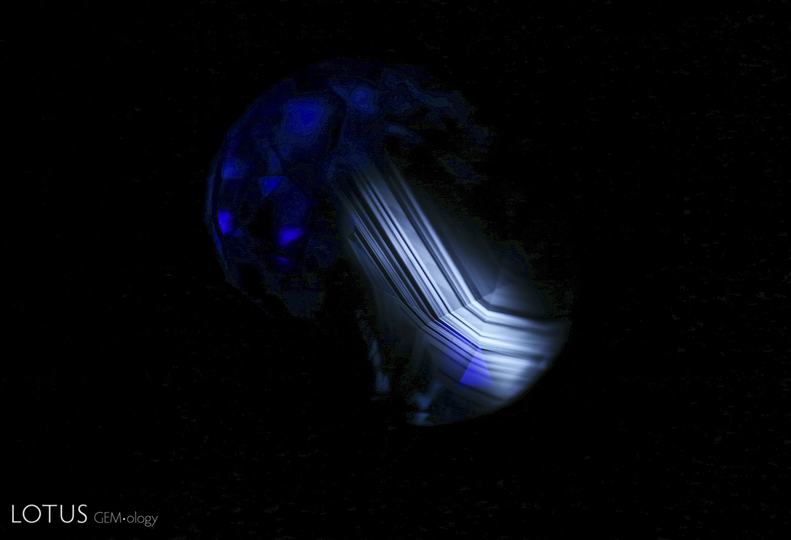
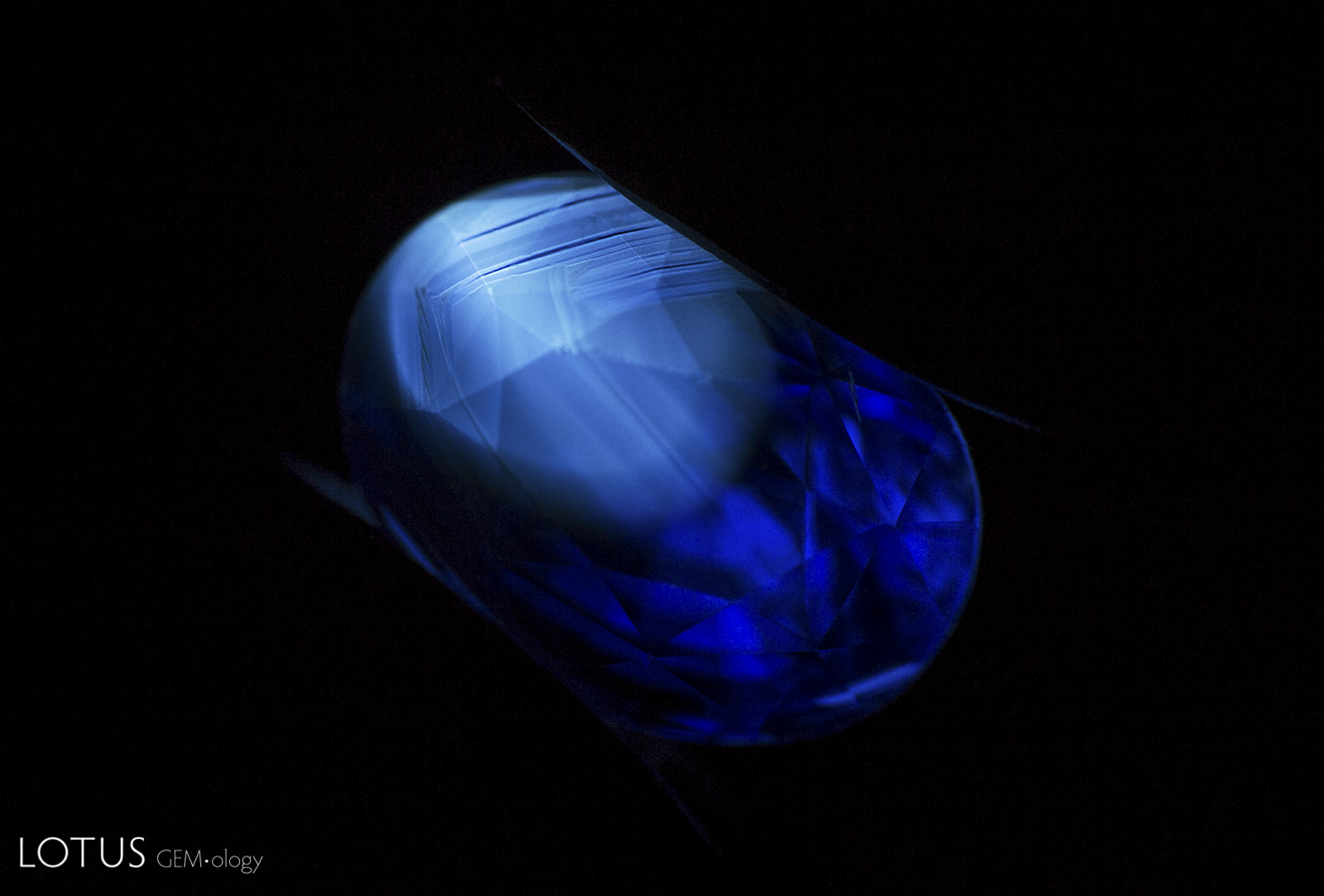
Angular zoned chalky shortwave fluorescence provides evidence that this Madagascar sapphire was heated.
Natural Sapphire •
Madagascar •
Enhancements:
Heat (H) •
Lighting Conditions:
Ultraviolet: Shortwave
Photographer:
E. Billie Hughes •
Image Number:
A-002-5043-2
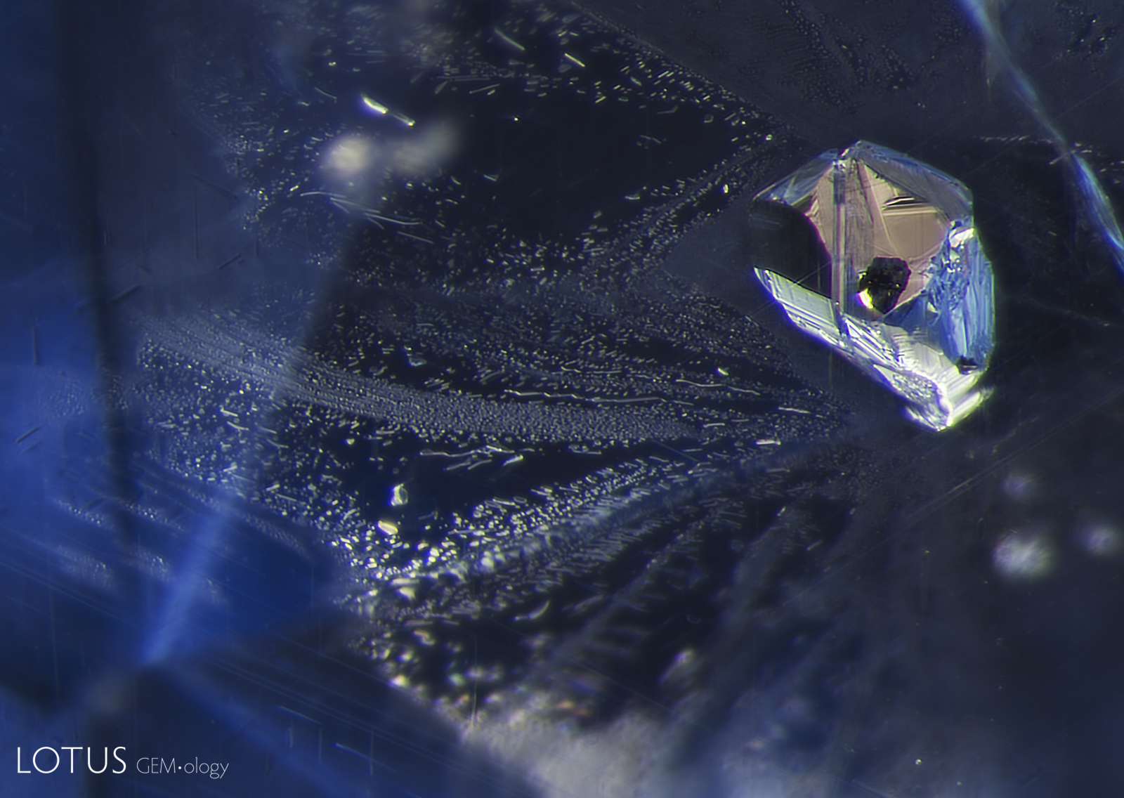
A small black crystal peeks out from inside a negative crystal where it is held inside a Sri Lankan sapphire.
Natural Sapphire •
Sri Lanka (Ceylon) •
Enhancements:
None Detected (None) •
Lighting Conditions:
Dark Field + Diffuse Fiber Optic
Photographer:
E. Billie Hughes •
Image Number:
A-002-4939-1
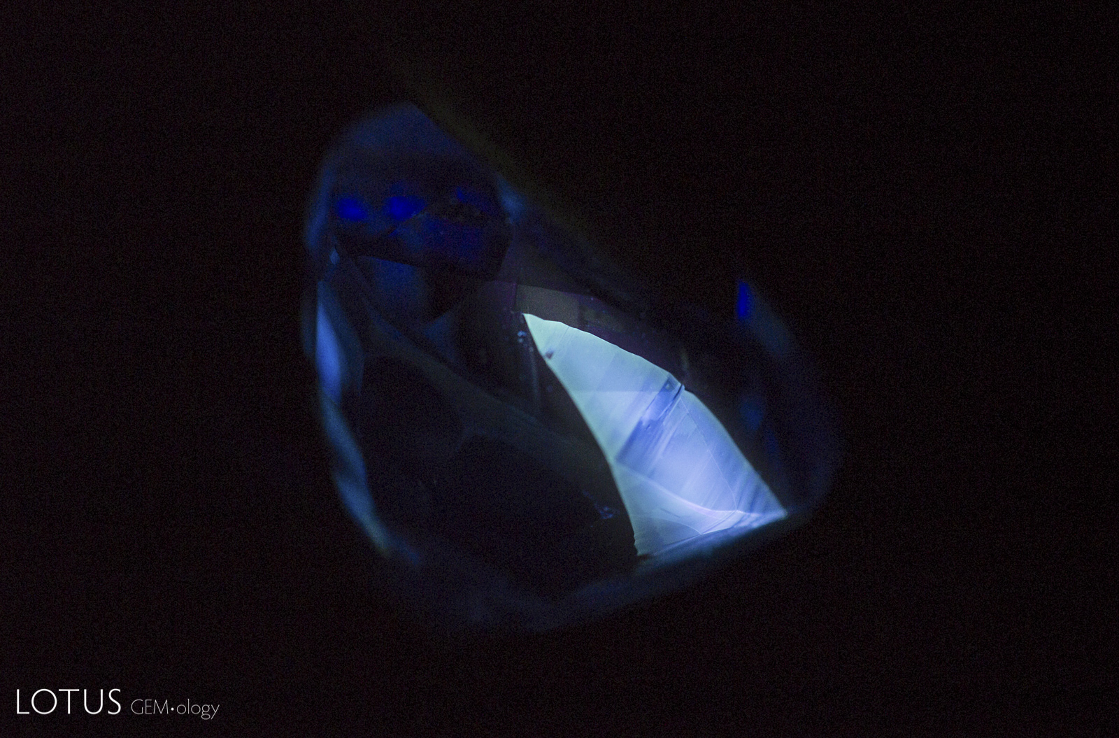
The angular chalky fluorescent patch in this sapphire provides evidence that the stone was heat treated.
Natural Sapphire •
Madagascar •
Enhancements:
Heat (H) •
Lighting Conditions:
Ultraviolet: Short Wave
Photographer:
E. Billie Hughes •
Image Number:
A-002-4843-2
This angular patch glows with a chalky appearance in shortwave UV light, providing evidence that the stone was heated.
Natural Sapphire •
Madagascar •
Enhancements:
Heat (H) •
Lighting Conditions:
Ultraviolet: Shortwave
Photographer:
E. Billie Hughes •
Image Number:
A-002-4783-1
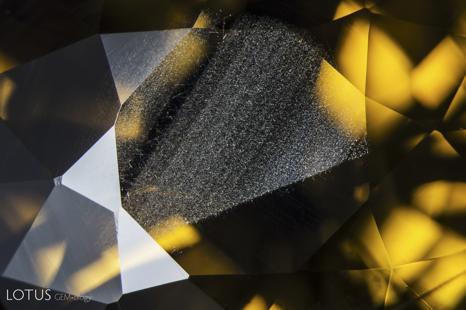
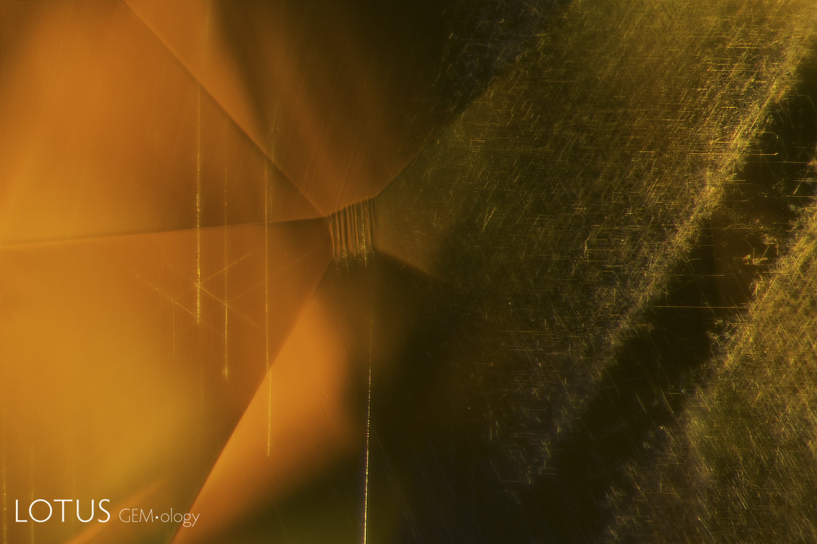
In yellow sapphire from Chanthaburi, Thailand, a thin layer of iron-rich silk is typically found in green layers on the skin of the crystal. After cutting, it is commonly seen just under the table and/or culet.
Natural Sapphire •
Thailand (Siam); Chanthaburi •
Enhancements:
None Detected (None) •
Lighting Conditions:
Fiber Optic: Diffuse Oblique
Photographer:
E. Billie Hughes •
Image Number:
A-002-4753-3
In yellow sapphire from Chanthaburi, Thailand, a thin layer of iron-rich silk is typically found in green layers on the skin of the crystal. After cutting, it is commonly seen just under the table and/or culet. In this example we can also see a series of fine boehmite needles on the left.
Natural Sapphire •
Thailand (Siam); Chanthaburi •
Enhancements:
None Detected (None) •
Lighting Conditions:
Dark Field + Shadowing
Photographer:
E. Billie Hughes •
Image Number:
A-002-4753-1
The shiny, melted appearance of this crystal and the fingerprint surrounding it suggest that their Madagascar sapphire host was heated.
Natural Sapphire •
Madagascar •
Enhancements:
Heat (H) •
Lighting Conditions:
Dark Field + Diffuse Fiber Optic + Shadowing
Photographer:
E. Billie Hughes •
Image Number:
A-002-4731-1
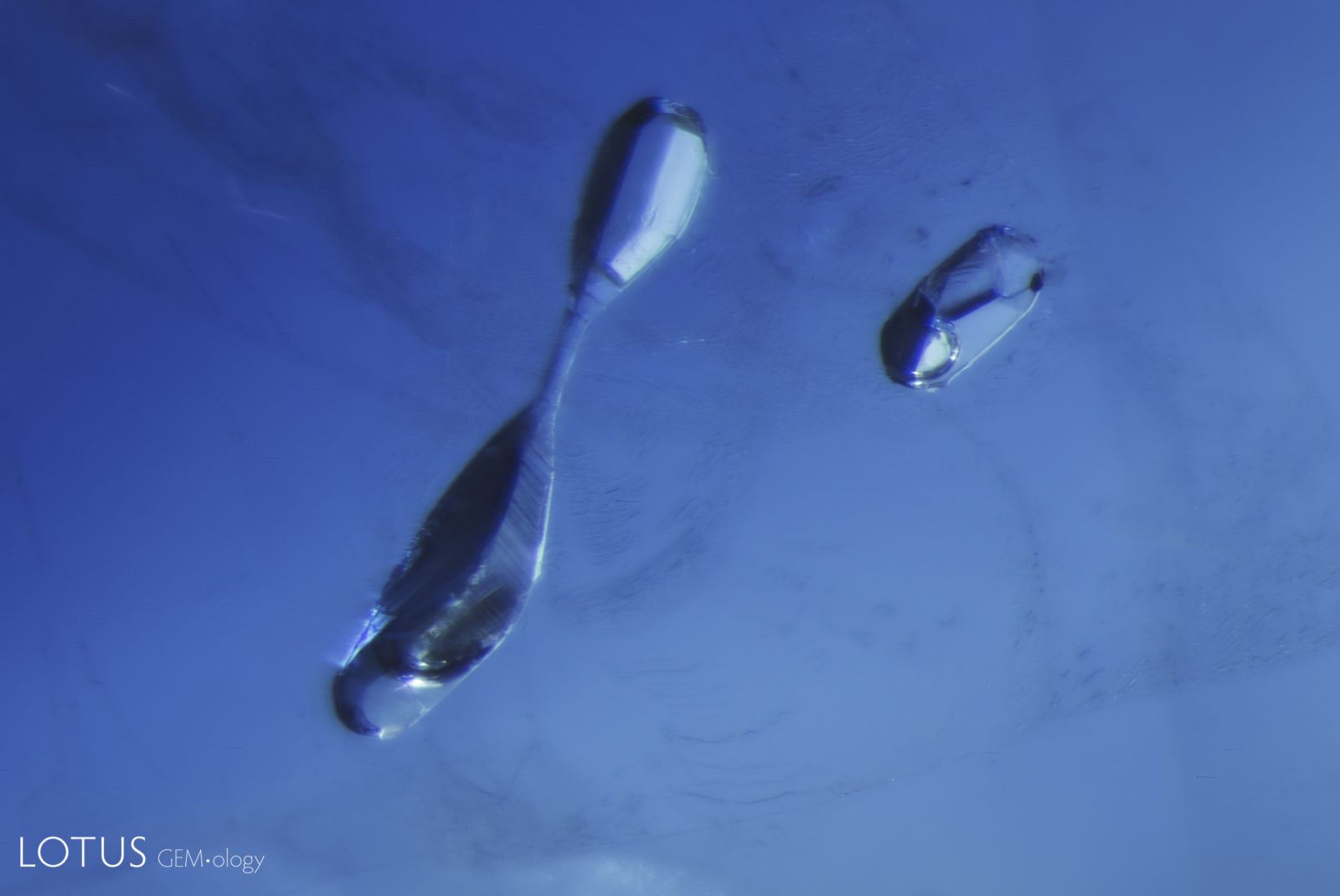
Small “frozen” gas bubbles trapped in place inside negative crystals provide evidence that a stone has been heated, as seen in this example in a basalt-related sapphire.
Natural Sapphire •
Basaltic •
Enhancements:
Heat (H) •
Lighting Conditions:
Light Field + Oblique Fiber Optic
Photographer:
E. Billie Hughes •
Image Number:
A-002-4703-3
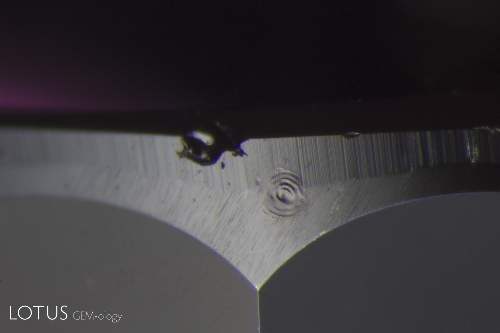
Laser-Induced-Breakdown-Spectroscopy (LIBS) is used by some gem labs to test for beryllium. Unlike Laser Ablation Inductively Coupled Plasma Mass Spectroscopy (LA-ICPMS), the surface is actually melted (rather than ablated), producing the circular rippled mark we see in the center of the girdle on this stone.
Natural Sapphire •
Madagascar •
Enhancements:
Heat (H) •
Lighting Conditions:
Diffuse Overhead
Photographer:
Richard W. Hughes •
Image Number:
A-002-4711-1
Undissolved rutile silk intersects under the table of this untreated Sri Lanka sapphire.
Natural Sapphire •
Sri Lanka (Ceylon) •
Enhancements:
None Detected (None) •
Lighting Conditions:
Fiber Optic: Diffuse Oblique
Photographer:
E. Billie Hughes •
Image Number:
A-002-4456-2
Undissolved rutile silk intersects under the table of this untreated Sri Lanka sapphire.
Natural Sapphire •
Sri Lanka (Ceylon) •
Enhancements:
None Detected (None) •
Lighting Conditions:
Fiber Optic: Diffuse Oblique
Photographer:
E. Billie Hughes •
Image Number:
A-002-4456-1
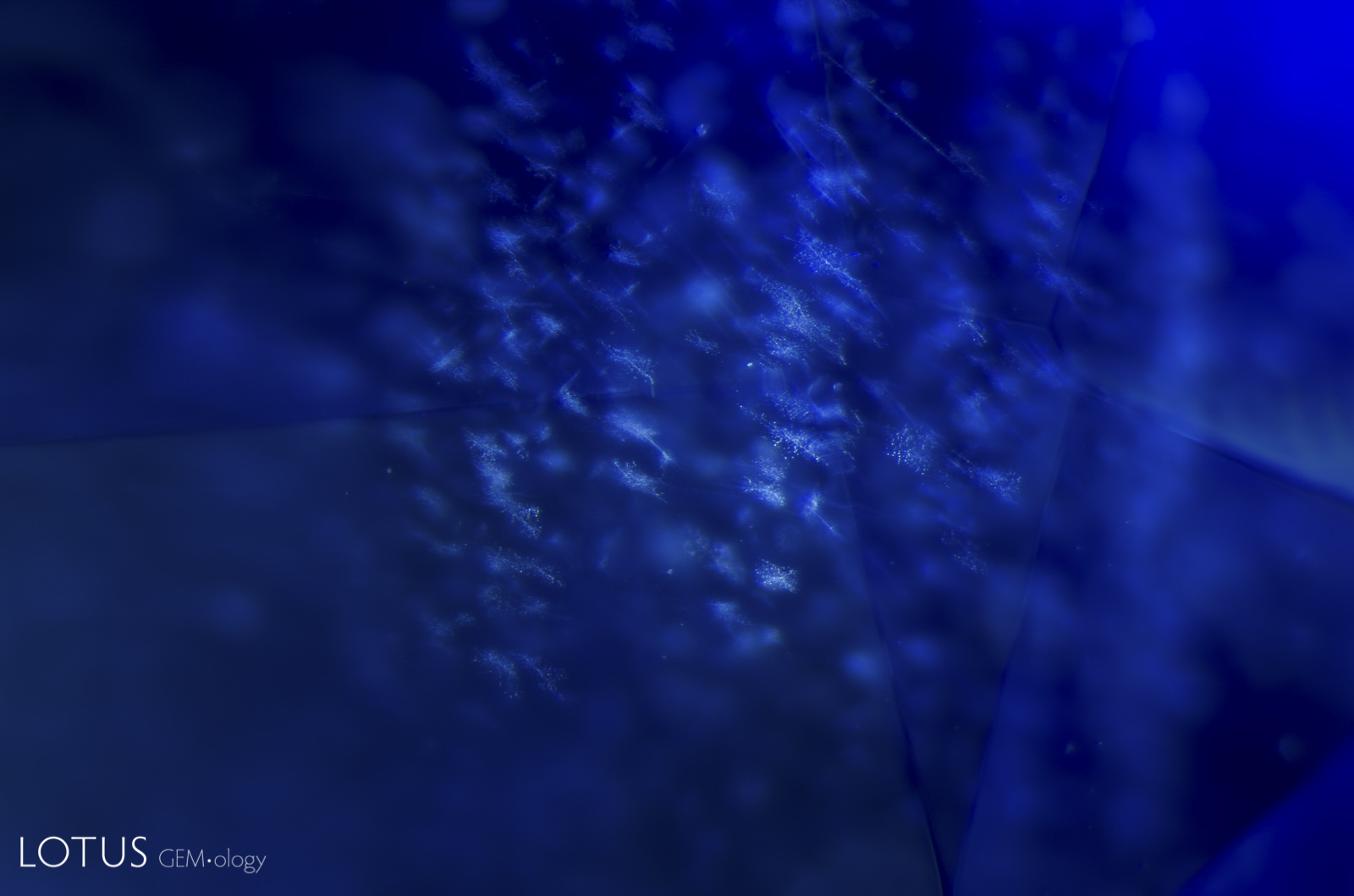
Small snowflake-like inclusions are a common feature in untreated Madagascar sapphire.
Natural Sapphire •
Madagascar •
Enhancements:
None Detected (None) •
Lighting Conditions:
Fiber Optic: Oblique
Photographer:
E. Billie Hughes •
Image Number:
A-002-4410-5
Sapphire inclusions may include mineral crystals, rutile silk, healed fissures, fluid inclusions, negative crystals, color zoning and other features formed during or after crystal growth. Such features are important to gemologists because they can provide evidence useful in identifying sapphire, understanding its geological history, recognizing treatment and, in some cases, determining geographic origin.
Sapphire Inclusion Photomicrographs
The Hyperion sapphire gallery includes microscopic features found in sapphires from major deposits around the world. Each entry is accompanied by descriptive information and, where available, references to the gemological literature.
What Inclusions Can Reveal About Sapphire
Inclusions are an important part of sapphire identification. Their form, composition, orientation and alteration can help gemologists distinguish natural sapphire from synthetic material, recognize evidence of heat treatment, and study the geological environment in which the sapphire formed.
Hyperion combines inclusion photomicrography with supporting gemological information so that individual features can be compared with documented examples from known sapphire deposits.
About Hyperion
Hyperion is the Lotus Gemology searchable inclusion database. It allows users to browse gemstone inclusions by gem type, geographic origin, treatment and keyword.