Treated Sapphire Inclusions
Explore treated sapphire inclusions in Hyperion, the Lotus Gemology inclusion database. Hyperion contains photomicrographs of inclusions and internal features in natural sapphires that have undergone treatment, documented with information on geographic origin, treatment, lighting conditions, field of view, photographer and published references.
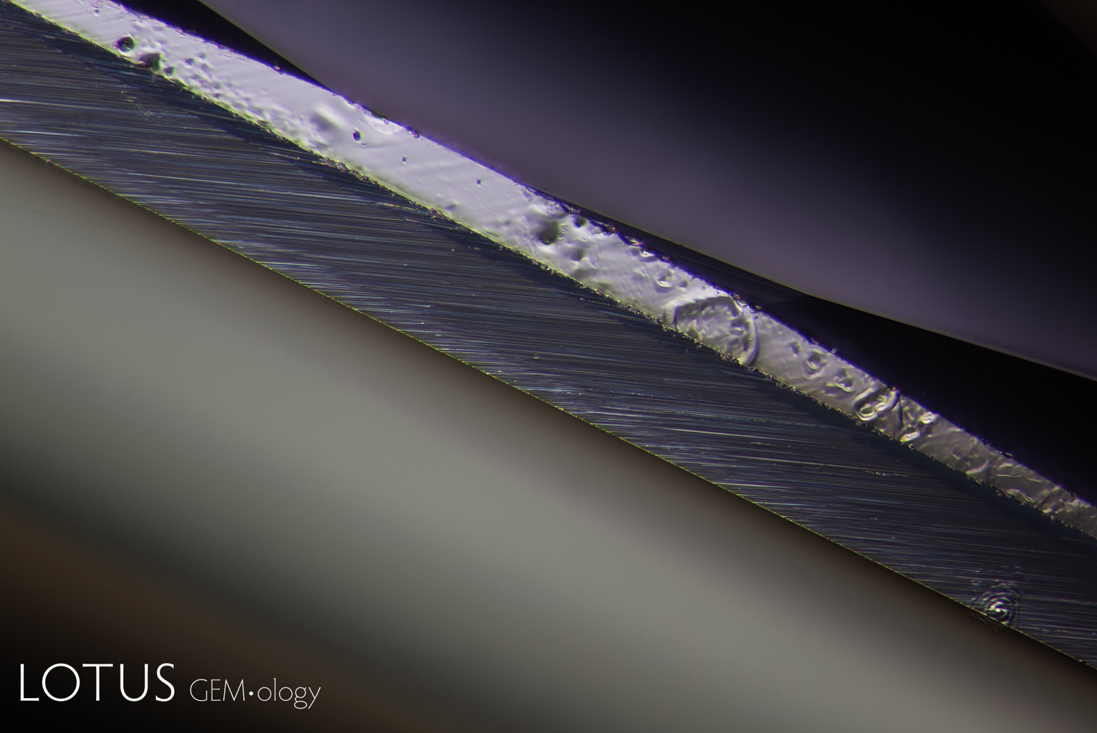
The high temperature that this blue sapphire from Madagascar was exposed to during beryllium diffusion treatment melted part of the girdle of the stone. Also, in the lower right corner of the photo is the tiny ablation spot that the LIBS created in order to detect the beryllium.
Natural Sapphire •
Madagascar •
Enhancements:
Heat + Diffusion of external coloring agents (H-D) •
Lighting Conditions:
Diffuse Dark Field with Blue Filter + Diffuse Fibre Optic
Photographer:
Kaylan Khourie •
Image Number:
A-005-2634-3
• Field of View = 2.8mm
Hughes, R.W., Manorotkul, W. and Hughes, E.B. (2017) Ruby & Sapphire: A Gemologist's Guide. Bangkok, Lotus Publishing, 816 pp.; RWHL*.
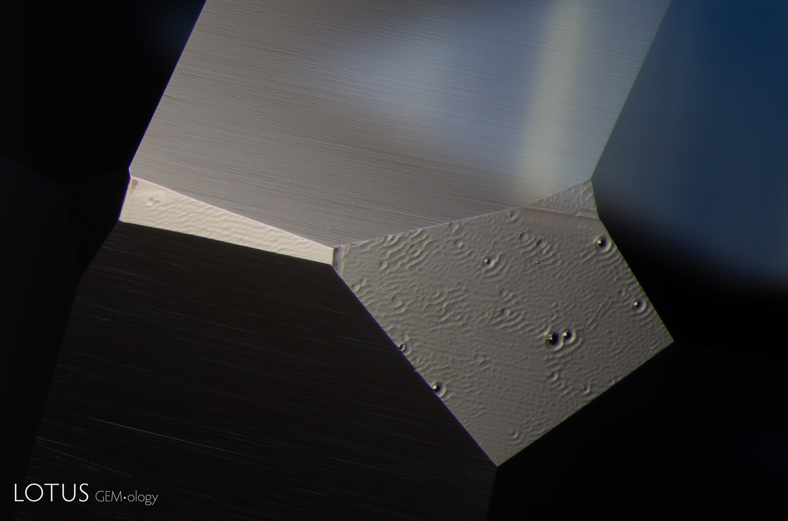
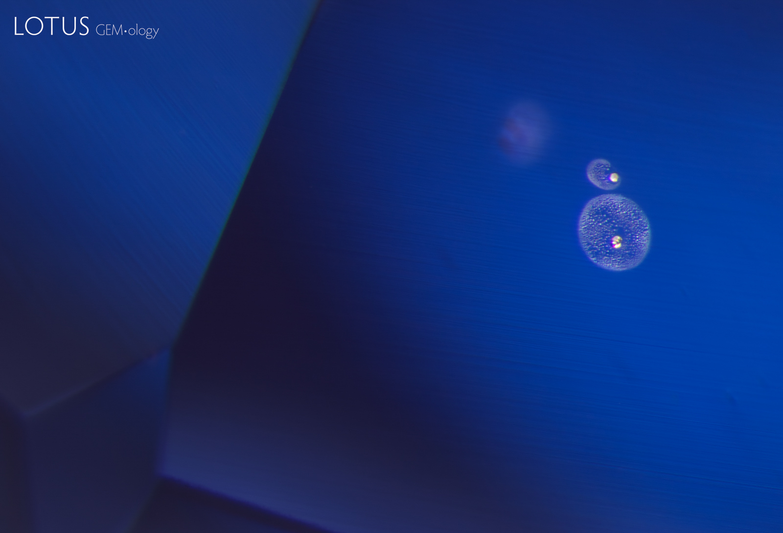
A subtle sign of treatment. Ruby and sapphire surfaces often develop a melted texture when heated, sometimes forming “frozen” droplets that resemble water but cannot be wiped away. To minimize this appearance, stones are frequently repolished. In this case, a few facets were missed, leaving behind telltale signs of heat treatment.
Natural Sapphire •
Sri Lanka (Ceylon) •
Enhancements:
Heat (H) •
Lighting Conditions:
Dark Field + Overhead Fiber Optic
Photographer:
E. Billie Hughes •
Image Number:
A-005-6905-1
• Field of View = 4mm
Hughes, R.W., Manorotkul, W. and Hughes, E.B. (2017) Ruby & Sapphire: A Gemologist's Guide. Bangkok, Lotus Publishing, 816 pp.; RWHL*.
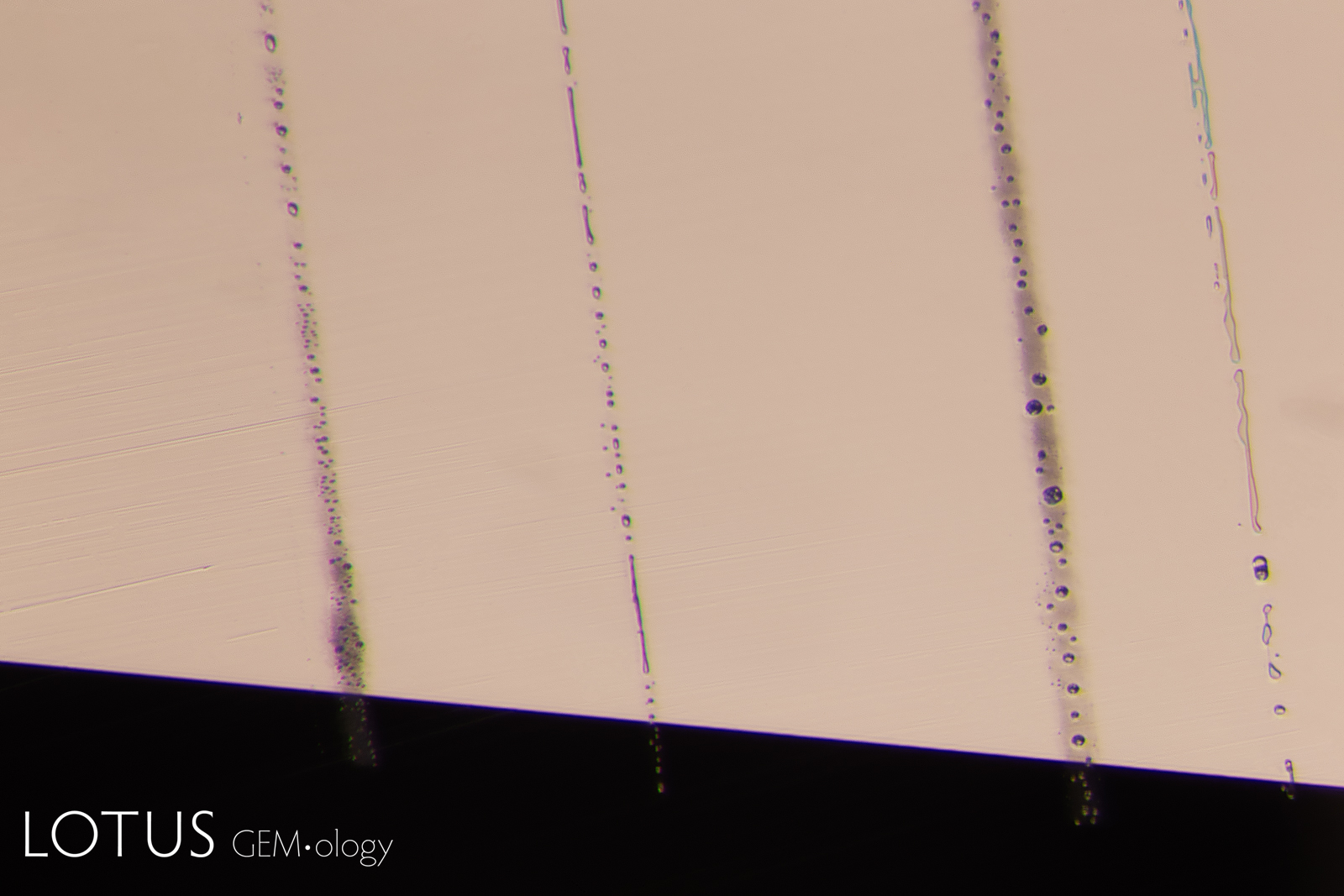
These dotted outlines, sometimes called “crop circles,” can occasionally be found in blue sapphires heated at high temperatures. While frequently associated with beryllium diffused stones, they are not conclusive evidence of diffusion treatment. The stone pictured here was heated, but showed no evidence of beryllium diffusion.
Natural Sapphire •
Sri Lanka (Ceylon) •
Enhancements:
Heat (H) •
Lighting Conditions:
Dark Field + Oblique Fiber Optic
Photographer:
E. Billie Hughes •
Image Number:
A-005-6689-1
• Field of View = 2mm
Hughes, R.W., Manorotkul, W. and Hughes, E.B. (2017) Ruby & Sapphire: A Gemologist's Guide. Bangkok, Lotus Publishing, 816 pp.; RWHL*.
Du Toit, G., Hughes, R.W. et al. (2006) Beryllium-treated blue sapphires. Bangkok, www.ruby-sapphire.com, RWHL.
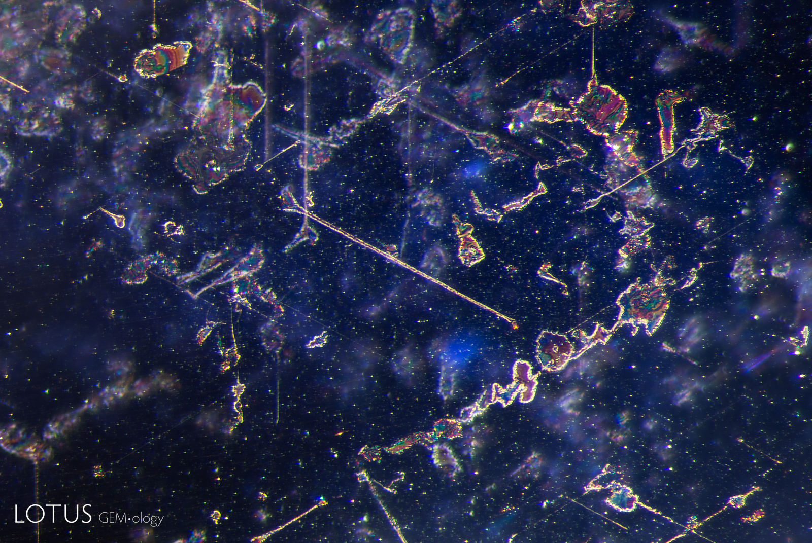
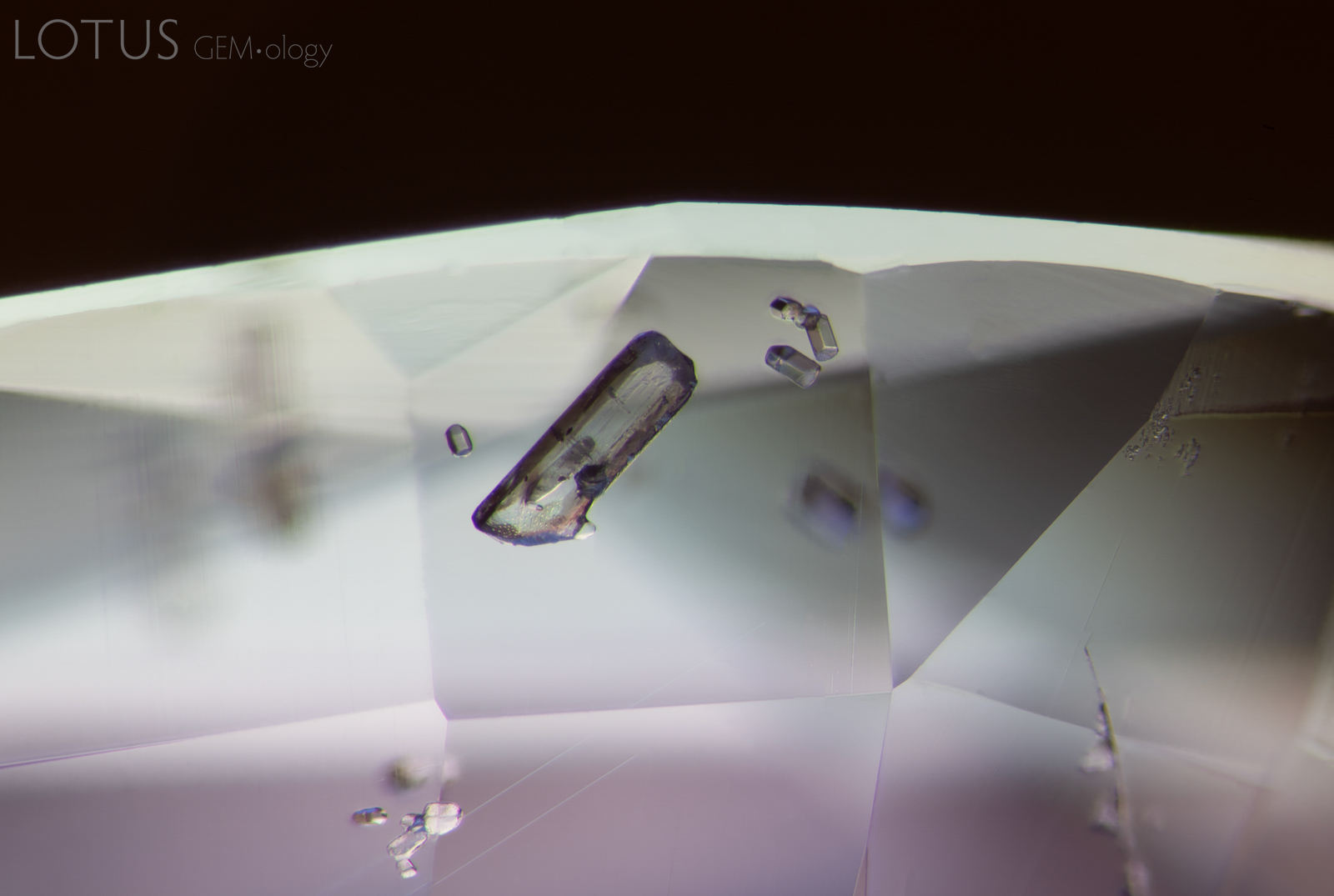
This blue sapphire from Myanmar’s Mogok Valley contains several prismatic apatite crystals. Observation between crossed polarized filters reveals not only the bright interference colors of the apatite, but also of its sub-inclusions.
Natural Sapphire •
Myanmar (Burma); Mogok/Namya •
Enhancements:
Fissure Filling with colorless Oil/resin (FF-O1); No indications of heating (No indications of heating) •
Lighting Conditions:
Dark Field + Crossed Polars
Photographer:
Kaylan Khourie •
Image Number:
A-005-4459-1
• Field of View = 3.6mm
Hughes, R.W., Manorotkul, W. and Hughes, E.B. (2017) Ruby & Sapphire: A Gemologist's Guide. Bangkok, Lotus Publishing, 816 pp.; RWHL*
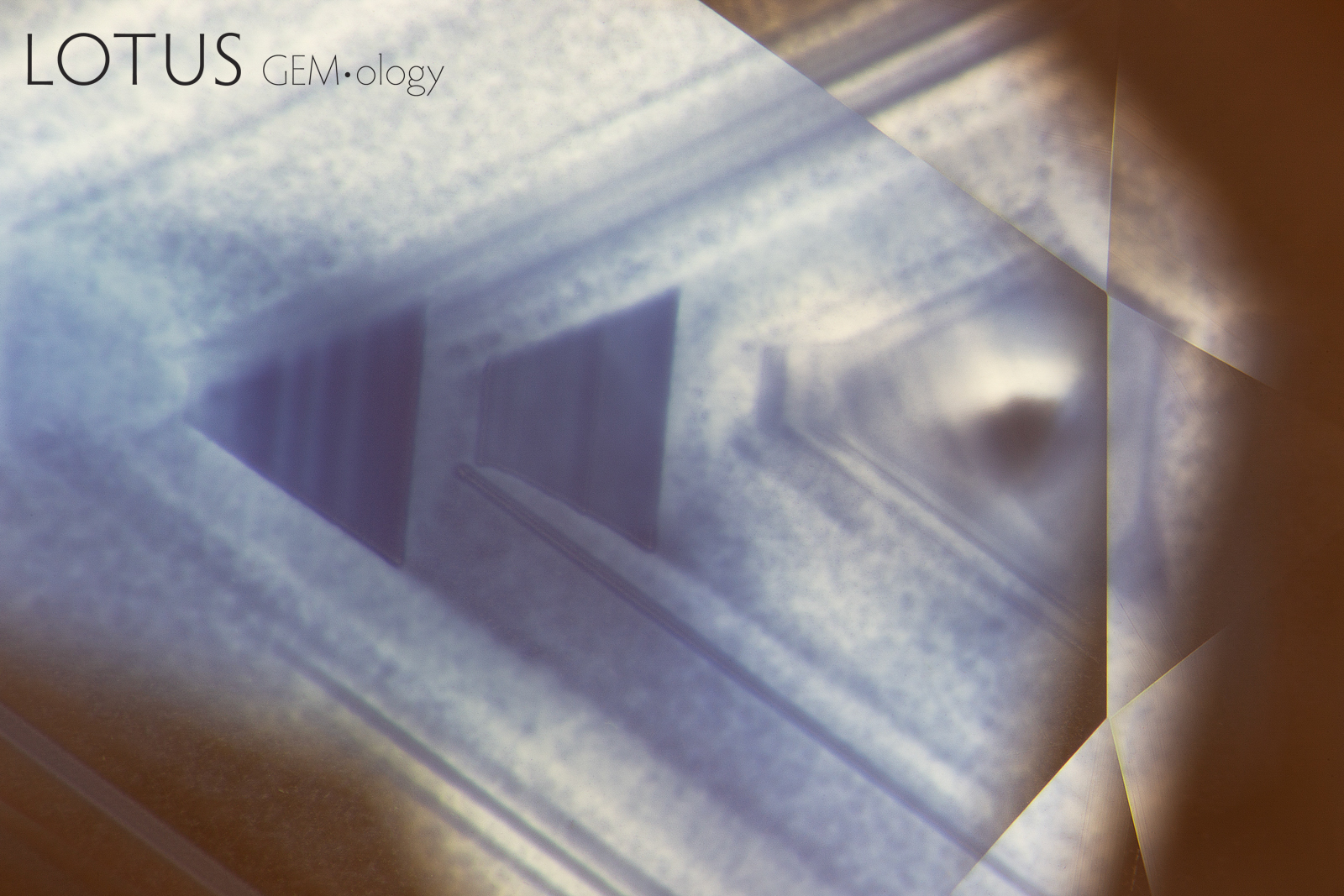
This high temperature heat-treated blue sapphire from a basalt-related source displays zoned internal diffusion. The high temperature (usually above 1600˚C) causes the rutile needles to dissolve and release titanium into the host sapphire, the titanium pairs with the iron in the stone and this creates blue color around the rutile silk remnants.
Natural Sapphire •
Basalt Related •
Enhancements:
Heat (H) •
Lighting Conditions:
Dark Field
Photographer:
Kaylan Khourie •
Image Number:
A-005-2573-1
• Field of View = 3.7mm
• Magnification = 4.8x
Hughes, R.W., Manorotkul, W. and Hughes, E.B. (2017) Ruby & Sapphire: A Gemologist's Guide. Bangkok, Lotus Publishing, 816 pp.; RWHL*.
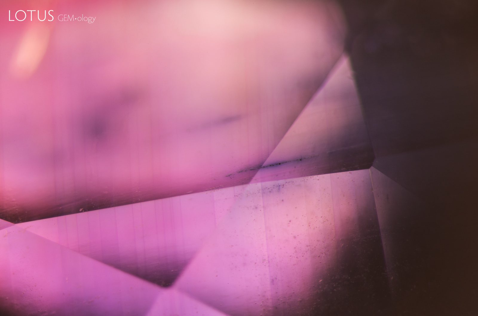
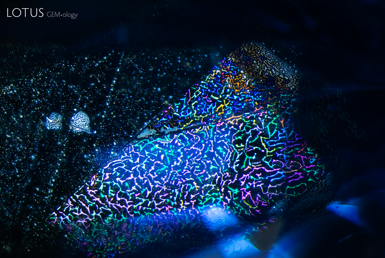
High temperature heat treatment melted the surface of this sapphire, resulting in recrystallized corundum at and just below the surface.
Natural Sapphire •
Basalt Related •
Enhancements:
Heat + Diffusion of external coloring agents (H-D) •
Lighting Conditions:
Light Field (Transmitted Light)
Photographer:
E. Billie Hughes •
Image Number:
A-005-1560-1
• Field of View = 7.2mm
Hughes, R.W., Manorotkul, W. and Hughes, E.B. (2017) Ruby & Sapphire: A Gemologist's Guide. Bangkok, Lotus Publishing, 816 pp.; RWHL*.
Emmett, J.L., Scarratt, K. et al. (2003) Beryllium diffusion of ruby and sapphire. Gems & Gemology, Vol. 39, No. 2, Summer, pp. 84–135; RWHL*.
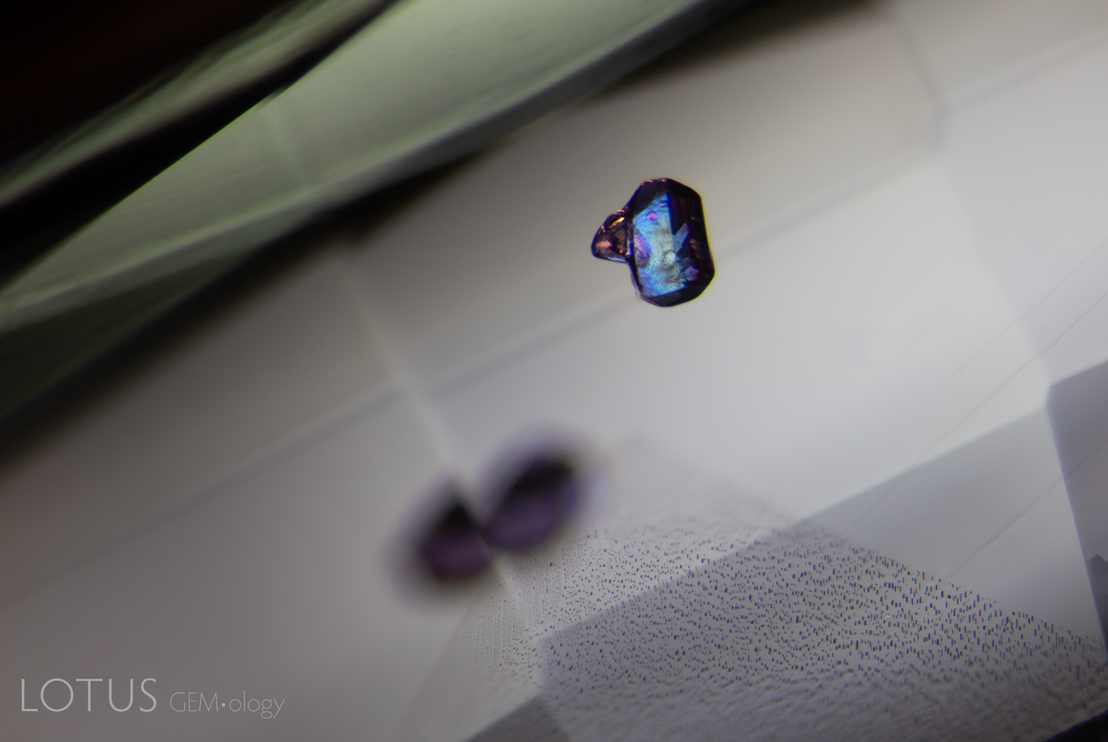
Some of the small particles in this sapphire are surrounded blue color concentrations. Often termed “internal diffusion,” this occurs when titanium from rutile dissolves (enters into solid solution) into the surrounding sapphire as a result of heat treatment. Once in solid solution, the Ti atoms combine with Fe atoms, producing an intervalence charge transfer that absorbs light.
Natural Sapphire •
Pakistan (Kashmir); Batakundi •
Enhancements:
Heat (H) •
Lighting Conditions:
Diffuse Light Field (Transmitted Light)
Photographer:
E. Billie Hughes •
Image Number:
A-005-1643-1
• Field of View = 5.1mm
Hughes, R.W., Manorotkul, W. and Hughes, E.B. (2017) Ruby & Sapphire: A Gemologist's Guide. Bangkok, Lotus Publishing, 816 pp.; RWHL*.
249 Total Images
Results: 13 – 24 | Page 2 of 21 Pages
Treatment can alter existing inclusions and produce new microscopic features in sapphire. These may include changes to mineral inclusions, altered rutile silk, tension fractures, healed fissures, residues and other features associated with heating, diffusion, fissure filling and other treatment processes.
Treated Sapphire Inclusion Photomicrographs
The Hyperion treated sapphire gallery includes microscopic features documented in natural sapphires subjected to treatment. Each entry identifies the recorded treatment and is accompanied by descriptive information and, where available, references to the gemological literature.
What Inclusions Can Reveal About Sapphire Treatment
Microscopic examination is an important part of detecting treatment in sapphire. The appearance and alteration of inclusions, fractures and growth features can provide evidence of treatment and, in some cases, information about the processes to which a sapphire has been subjected.
Hyperion combines inclusion photomicrography with supporting gemological information so that treatment-related features can be compared with documented examples in natural sapphire.
About Hyperion
Hyperion is the Lotus Gemology searchable inclusion database. It allows users to browse gemstone inclusions by gem type, geographic origin, treatment and keyword.
Search the Complete Hyperion Database