Treated Sapphire Inclusions
Explore treated sapphire inclusions in Hyperion, the Lotus Gemology inclusion database. Hyperion contains photomicrographs of inclusions and internal features in natural sapphires that have undergone treatment, documented with information on geographic origin, treatment, lighting conditions, field of view, photographer and published references.
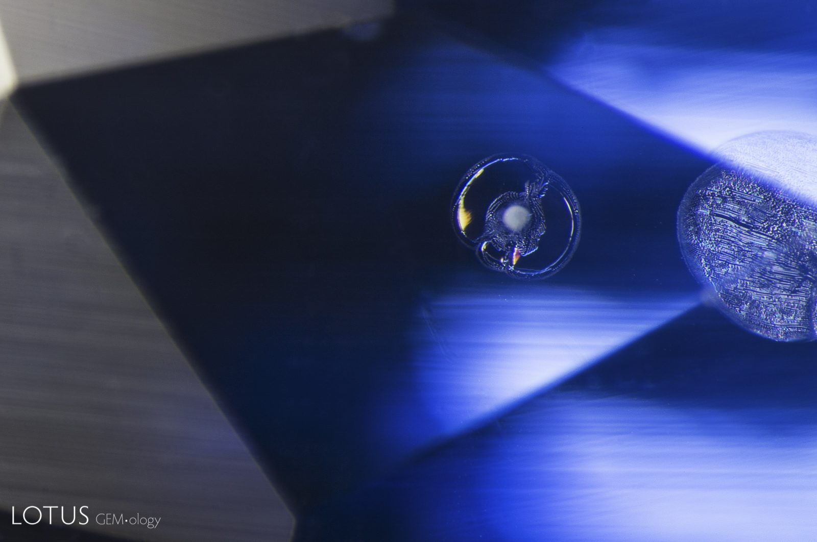
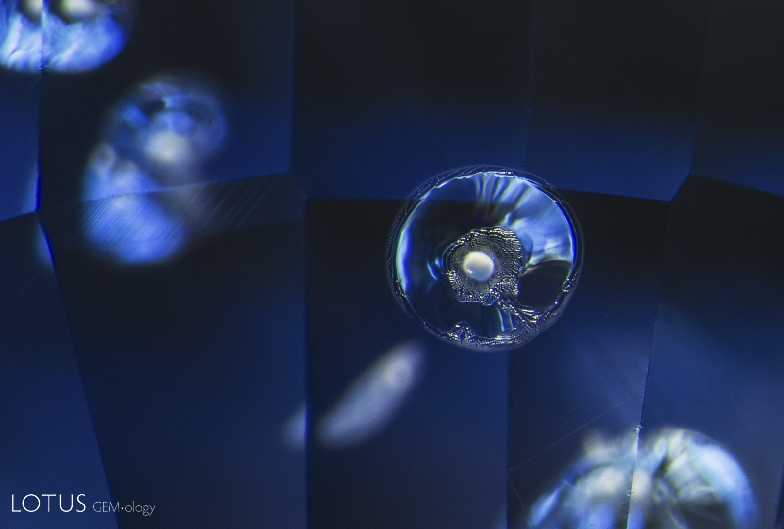
In this sapphire from Sri Lanka, evidence of high temperature heat treatment can be found in this moiré-patterned fingerprint. The once-lovely lacy pattern of liquid droplets is now besmirched by circular “explosions,” where the pressure from heating caused ruptures in the microscopic negative crystals, thus deflowering Mother Nature’s exquisite work of art.
Natural Sapphire •
Sri Lanka (Ceylon) •
Enhancements:
Heat (H) •
Lighting Conditions:
Dark Field + Shadowing
Photographer:
Richard W. Hughes •
Image Number:
A-002-6297-1
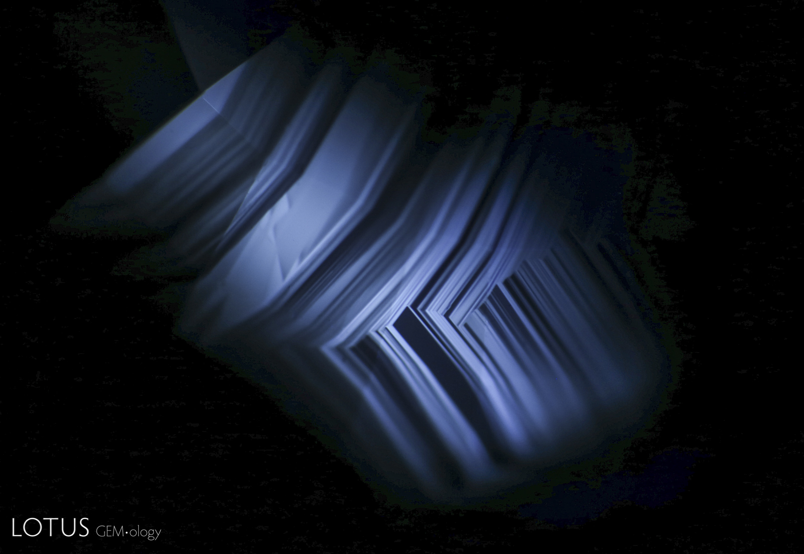
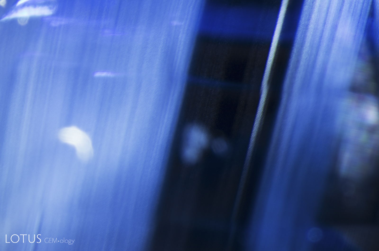
Angular zoned chalky shortwave fluorescence provides evidence that this Madagascar sapphire was heated.
Natural Sapphire •
Madagascar •
Enhancements:
Heat (H) •
Lighting Conditions:
Ultraviolet: Shortwave
Photographer:
E. Billie Hughes •
Image Number:
A-002-5043-3
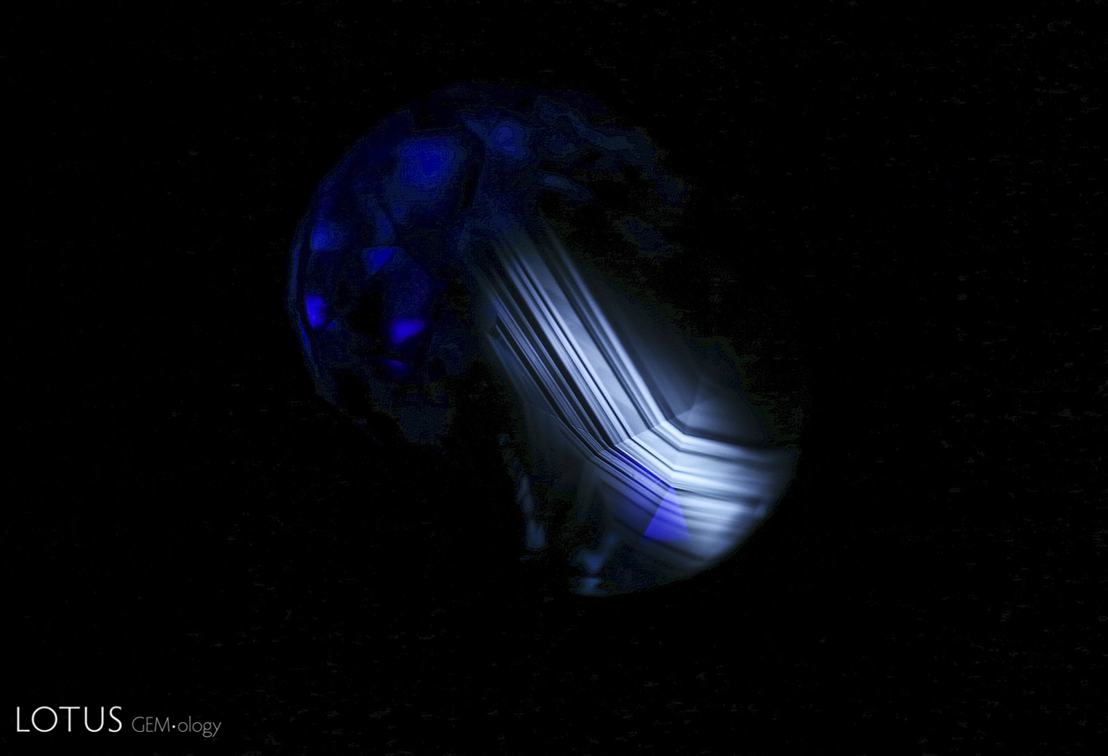
Angular zoned chalky shortwave fluorescence provides evidence that this Madagascar sapphire was heated.
Natural Sapphire •
Madagascar •
Enhancements:
Heat (H) •
Lighting Conditions:
Ultraviolet: Shortwave
Photographer:
E. Billie Hughes •
Image Number:
A-002-5043-2
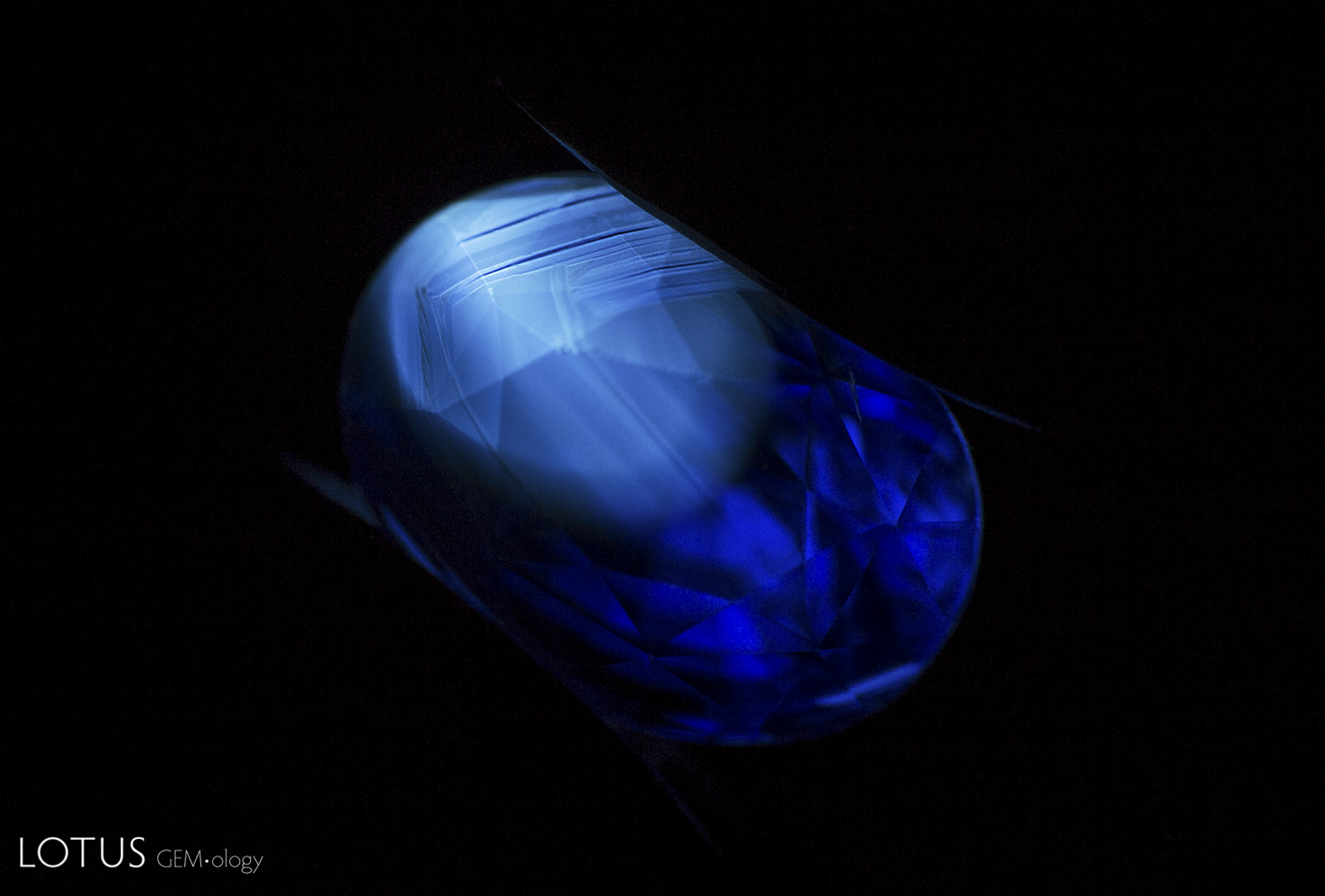
The angular chalky fluorescent patch in this sapphire provides evidence that the stone was heat treated.
Natural Sapphire •
Madagascar •
Enhancements:
Heat (H) •
Lighting Conditions:
Ultraviolet: Short Wave
Photographer:
E. Billie Hughes •
Image Number:
A-002-4843-2
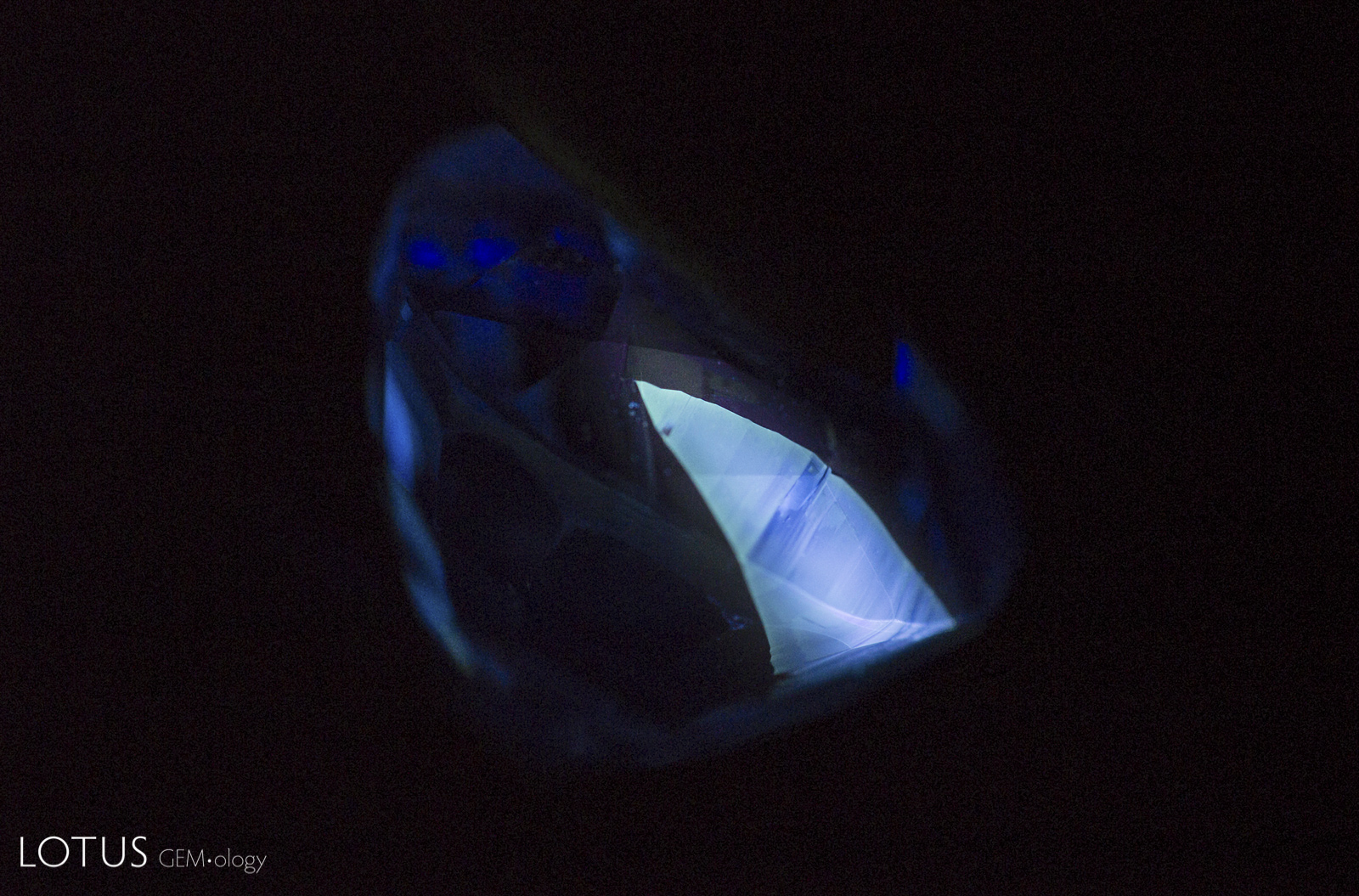
This angular patch glows with a chalky appearance in shortwave UV light, providing evidence that the stone was heated.
Natural Sapphire •
Madagascar •
Enhancements:
Heat (H) •
Lighting Conditions:
Ultraviolet: Shortwave
Photographer:
E. Billie Hughes •
Image Number:
A-002-4783-1
The shiny, melted appearance of this crystal and the fingerprint surrounding it suggest that their Madagascar sapphire host was heated.
Natural Sapphire •
Madagascar •
Enhancements:
Heat (H) •
Lighting Conditions:
Dark Field + Diffuse Fiber Optic + Shadowing
Photographer:
E. Billie Hughes •
Image Number:
A-002-4731-1
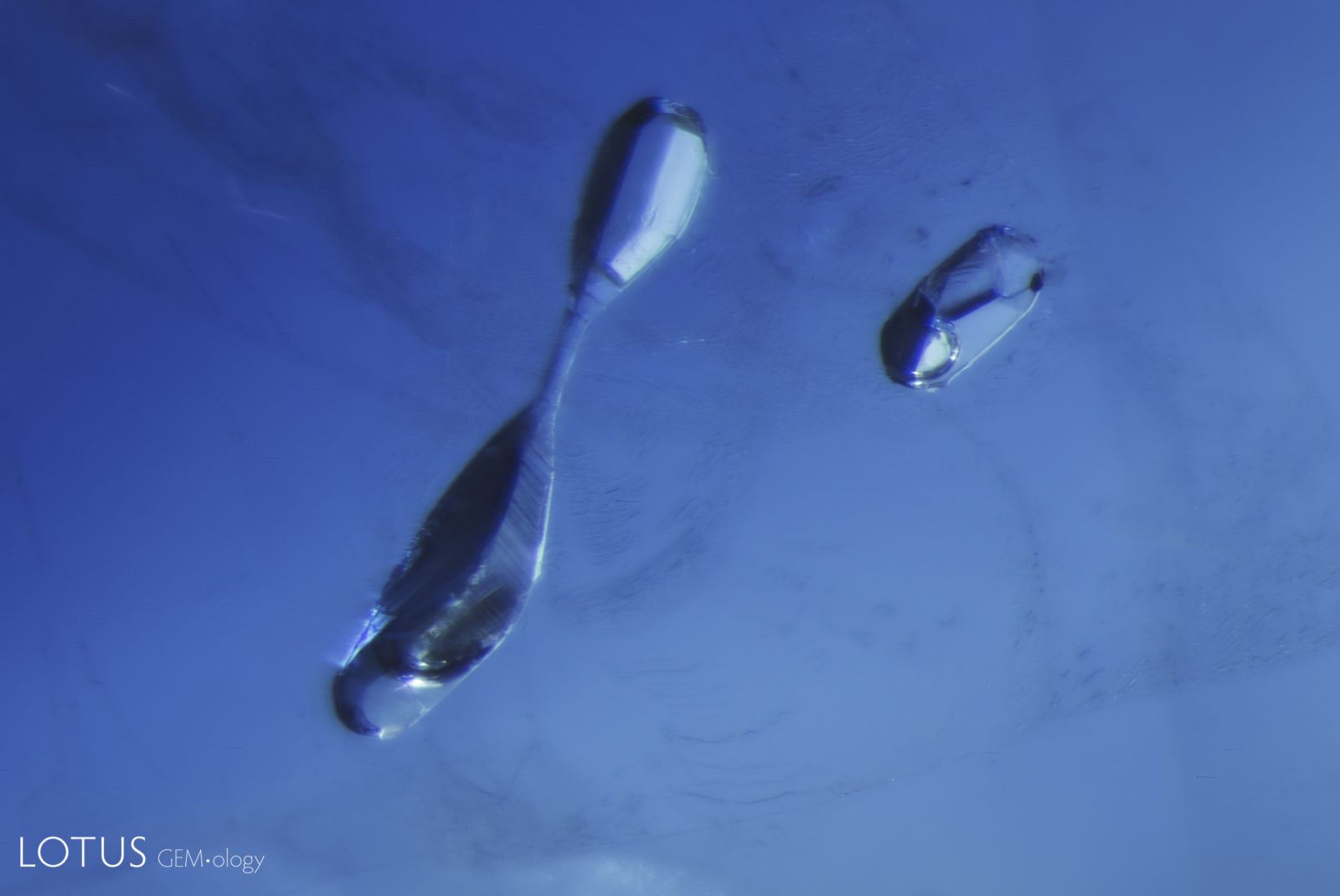
Small “frozen” gas bubbles trapped in place inside negative crystals provide evidence that a stone has been heated, as seen in this example in a basalt-related sapphire.
Natural Sapphire •
Basaltic •
Enhancements:
Heat (H) •
Lighting Conditions:
Light Field + Oblique Fiber Optic
Photographer:
E. Billie Hughes •
Image Number:
A-002-4703-3
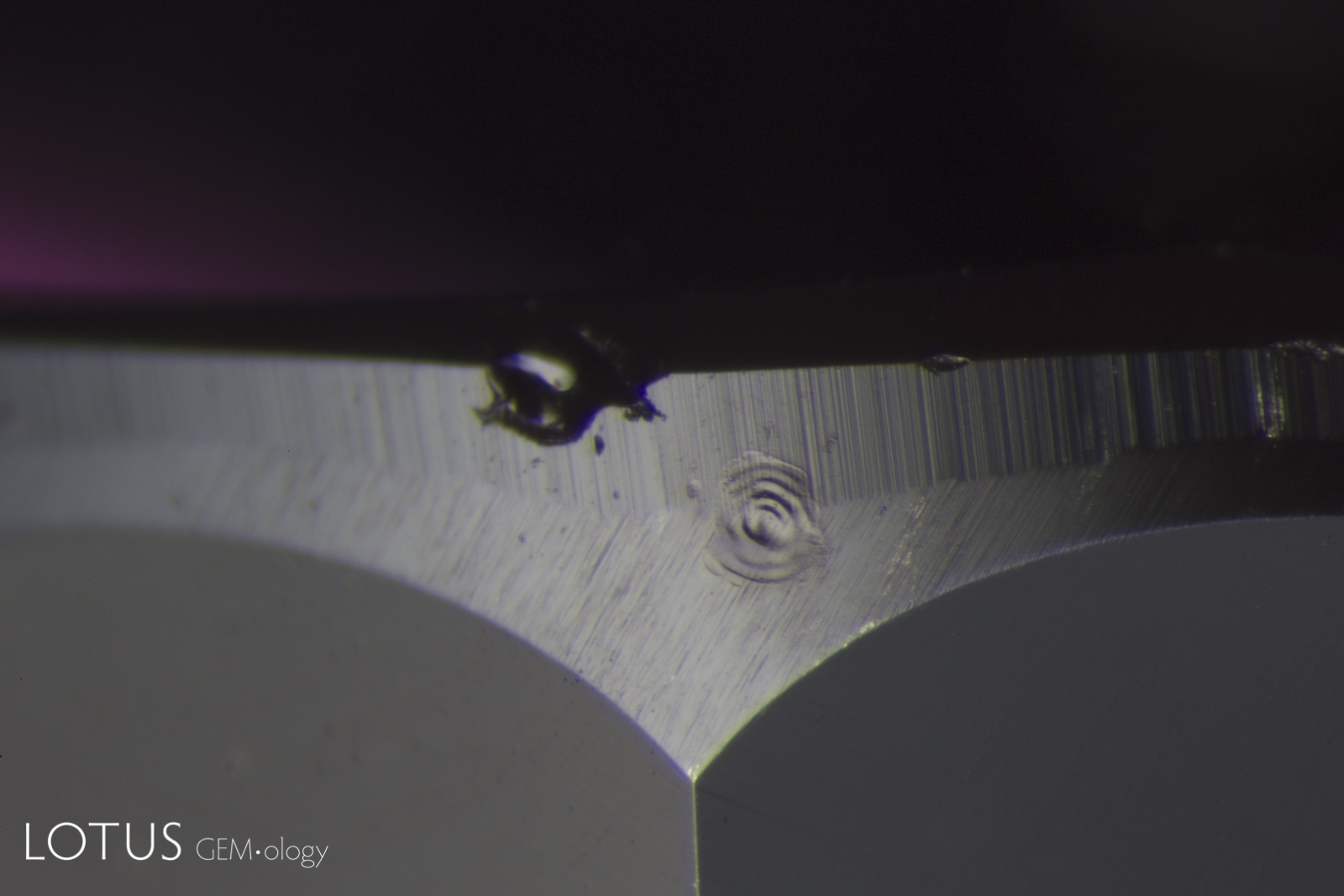
Laser-Induced-Breakdown-Spectroscopy (LIBS) is used by some gem labs to test for beryllium. Unlike Laser Ablation Inductively Coupled Plasma Mass Spectroscopy (LA-ICPMS), the surface is actually melted (rather than ablated), producing the circular rippled mark we see in the center of the girdle on this stone.
Natural Sapphire •
Madagascar •
Enhancements:
Heat (H) •
Lighting Conditions:
Diffuse Overhead
Photographer:
Richard W. Hughes •
Image Number:
A-002-4711-1
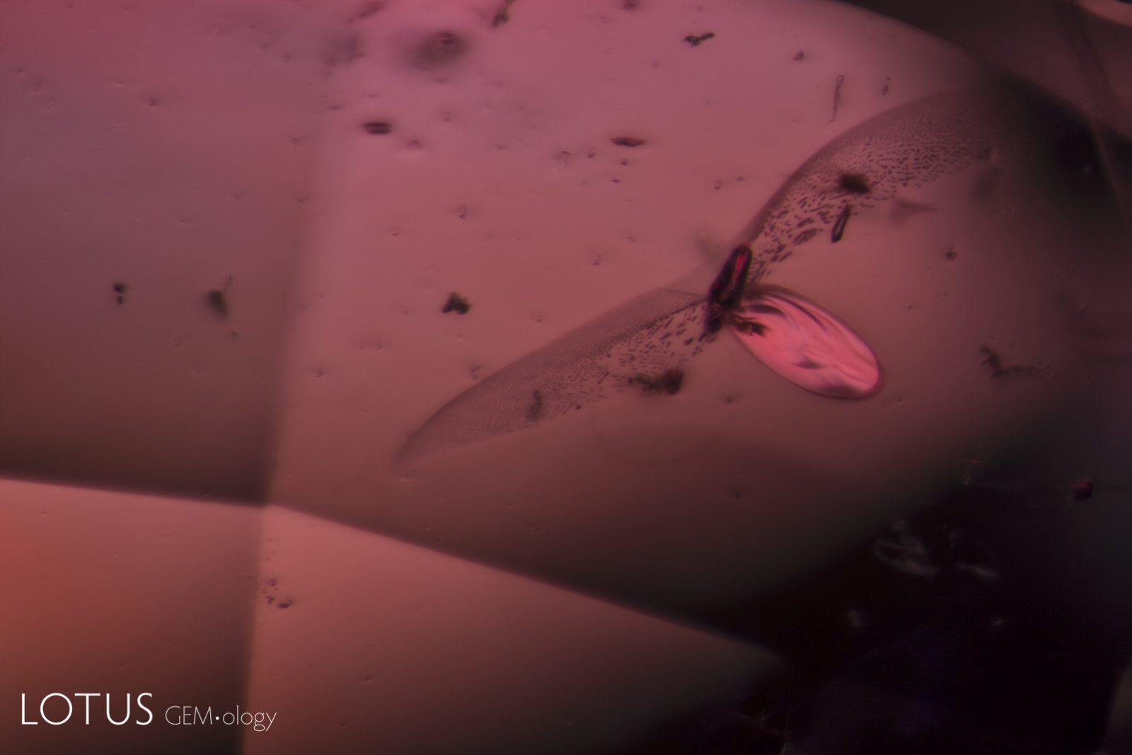
Resembling a large insect, this crystal with a discoid fissure was found in a heat-treated padparadscha sapphire from Madagascar.
Natural Padparadscha Sapphire •
Madagascar •
Enhancements:
Heat (H) •
Lighting Conditions:
Dark Field
Photographer:
Richard W. Hughes •
Image Number:
A-002-4444-1
Dense texture clouds made of tiny exsolved particles form angular zones in this heated Madagascar sapphire.
Natural Sapphire •
Madagascar •
Enhancements:
Heat (H) •
Lighting Conditions:
Fiber Optic: Oblique
Photographer:
E. Billie Hughes •
Image Number:
A-002-3764-1
Frosty melted “snowball” crystals provide evidence that this Madagascar sapphire was heat treated.
Natural Sapphire •
Madagascar •
Enhancements:
Heat (H) •
Lighting Conditions:
Fiber Optic: Diffuse Oblique
Photographer:
E. Billie Hughes •
Image Number:
A-002-3763-2
Frosty melted “snowball” crystals provide evidence that this Madagascar sapphire was heat treated.
Natural Sapphire •
Madagascar •
Enhancements:
Heat (H) •
Lighting Conditions:
Fiber Optic: Diffuse Oblique
Photographer:
E. Billie Hughes •
Image Number:
A-002-3763-1
Treatment can alter existing inclusions and produce new microscopic features in sapphire. These may include changes to mineral inclusions, altered rutile silk, tension fractures, healed fissures, residues and other features associated with heating, diffusion, fissure filling and other treatment processes.
Treated Sapphire Inclusion Photomicrographs
The Hyperion treated sapphire gallery includes microscopic features documented in natural sapphires subjected to treatment. Each entry identifies the recorded treatment and is accompanied by descriptive information and, where available, references to the gemological literature.
What Inclusions Can Reveal About Sapphire Treatment
Microscopic examination is an important part of detecting treatment in sapphire. The appearance and alteration of inclusions, fractures and growth features can provide evidence of treatment and, in some cases, information about the processes to which a sapphire has been subjected.
Hyperion combines inclusion photomicrography with supporting gemological information so that treatment-related features can be compared with documented examples in natural sapphire.
About Hyperion
Hyperion is the Lotus Gemology searchable inclusion database. It allows users to browse gemstone inclusions by gem type, geographic origin, treatment and keyword.